Chronic migraine & brain changes are directly connected. Chronic migraine is defined as 15 or more headache days per month for at least 3 months, with at least 8 meeting full migraine criteria. Research from the American Migraine Foundation confirms that repeated attacks alter brain structure, white matter integrity, and cognitive function over time.
Approximately 3.2 million US adults live with chronic migraine. This article covers how chronic migraines affect the brain, structural changes, memory effects, genetics, neuroinflammation, and evidence-based prevention strategies.
How Chronic Migraines Affect the Brain
How chronic migraines affect the brain goes far beyond pain during an attack. Each migraine triggers cortical spreading depression (CSD), a wave of intense electrical activity that rolls across the brain’s outer layer and temporarily shuts down normal function behind it.
During CSD, the trigeminal nerve releases calcitonin gene-related peptide (CGRP). CGRP sensitizes pain pathways. Each attack sensitizes them further. This is called central sensitization, and it is the core mechanism that converts episodic migraine into chronic migraine over years.
How chronic migraines affect the brain also includes measurable blood flow changes. MRI studies show reduced gray matter volume in the prefrontal cortex and somatosensory cortex of chronic migraine patients compared to headache-free controls. These regions control pain perception, decision-making, and sensory processing.
One attack leaves minimal lasting change. Fifteen-plus attacks per month, sustained over years, produce visible differences on brain imaging.
Brain Structure Changes in Chronic Migraine
Chronic migraine & brain changes visible on MRI fall into two main categories: white matter hyperintensities and gray matter volume loss.
White matter hyperintensities (WMH):
These appear as bright spots on MRI. They represent damaged nerve fiber insulation. A 2016 meta-analysis in Cephalalgia found migraine patients carry a 68% higher prevalence of WMH compared to headache-free controls. Migraine with aura accumulates these lesions faster than migraine without aura.
The posterior circulation territory (cerebellum and brainstem) collects WMH most frequently in migraine patients. This region supplies balance, coordination, and visual processing.
Gray matter changes:
A 2021 study in Brain found chronic migraine patients show measurable gray matter thinning in the orbitofrontal cortex after 5 years of chronic disease. The orbitofrontal cortex regulates pain expectation and emotional response to pain. Thinning here worsens pain catastrophizing, which amplifies perceived pain severity even outside attacks.
Memory Problems in Chronic Migraine
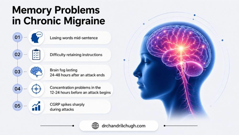
Memory problems chronic migraine patients experience are real and measurable. They are not simply a byproduct of pain or poor sleep.
A 2022 study in the Journal of Headache and Pain found chronic migraine patients scored significantly lower on verbal memory and processing speed tests compared to healthy controls, independent of depression, anxiety, or sleep quality scores.
The mechanism links directly to CGRP and the hippocampus. CGRP spikes sharply during attacks. In chronic migraine patients, baseline CGRP levels remain elevated between attacks. This sustained elevation creates persistent low-grade hippocampal impairment. The hippocampus is the brain’s primary memory-forming region.
Memory problems chronic migraine patients most commonly report:
- Losing words mid-sentence (the tip-of-tongue phenomenon)
- Difficulty retaining instructions or tasks within minutes
- Brain fog lasting 24-48 hours after an attack ends (the postdrome phase)
- Concentration problems in the 12-24 hours before an attack begins (the prodrome phase)
Memory problems chronic migraine patients experience are largely reversible with effective treatment. Clinical trials of anti-CGRP therapies like erenumab and fremanezumab show cognitive score improvement alongside reduced attack frequency.
Genetics and Chronic Migraine Risk
Familial hemiplegic migraine (FHM) is the clearest genetic form. It involves mutations in three genes: CACNA1A (calcium channel), ATP1A2 (sodium-potassium pump), and SCN1A (sodium channel). These mutations impair the brain’s ion flow regulation, making cortical spreading depression significantly easier to trigger.
For common chronic migraine, genome-wide association studies have identified 38+ genetic loci linked to migraine susceptibility. A 2022 study in Nature Genetics identified KCNK5 and TRPM8 gene variants as particularly strong risk factors for progression from episodic to chronic migraine.
Genetics and chronic migraine risk also interact with sex hormones. The MTHFR C677T gene variant impairs folate metabolism and raises homocysteine levels. Elevated homocysteine damages blood vessel linings and increases cortical spreading depression susceptibility. Women with this variant who use combined oral contraceptives face substantially elevated stroke risk alongside migraine with aura.
Genetics and chronic migraine risk do not make chronic migraine inevitable. Genetic predisposition requires environmental and lifestyle triggers to activate chronification. Reducing those triggers remains effective even in high-risk genetic profiles.
Triggers That Drive Brain Changes in Migraine
Chronic migraine & brain changes accelerate when specific triggers compound over time. Most patients focus on managing individual attacks. The more clinically significant question is what pushes the brain toward chronification.
Chronification triggers supported by research:
- Medication overuse : Using acute migraine medications (triptans, NSAIDs, opioids) on 10 or more days per month produces medication overuse headache (MOH). MOH restructures pain processing pathways and is the strongest modifiable driver of episodic-to-chronic conversion.
- Sleep disruption : Insomnia and untreated sleep apnea both increase attack frequency. The glymphatic system clears CGRP and inflammatory debris from brain tissue during sleep. Chronic poor sleep allows those compounds to accumulate.
- Obesity : Adipose tissue produces pro-inflammatory cytokines that raise baseline CGRP. A 2020 study in Neurology found obese episodic migraine patients were 81% more likely to develop chronic migraine within 11 years than normal-weight patients.
- Caffeine dependence : Regular high caffeine intake sensitizes adenosine receptors. Caffeine withdrawal between doses lowers the threshold for full migraine attacks through rebound headache.
- Hormonal fluctuation : Estrogen drops during menstruation, perimenopause, or contraceptive withdrawal trigger attacks consistently in susceptible women.
Neuroinflammation and Migraine Progression
Neuroinflammation drives chronic migraine & brain changes at the cellular level. It connects individual attacks to long-term brain alterations.
During migraine attacks, the trigeminal nucleus caudalis in the brainstem activates glial cells. These cells release IL-1β, TNF-alpha, and substance P. In episodic migraine, this inflammatory response resolves between attacks.
In chronic migraine, it does not fully resolve. Glial cell activation persists at a low level between attacks, a state called glial sensitization. This is responsible for the lowered pain threshold that makes chronic migraine patients sensitive to light, sound, and smell even between attacks.
Research published in Brain in 2023 confirmed elevated neuroinflammatory markers in the cerebrospinal fluid of chronic migraine patients that were absent in episodic migraine controls. Anti-CGRP monoclonal antibodies reduce this trigeminal neuroinflammation, which explains why they reduce attack frequency rather than only treating individual episodes.
Treatments to Prevent Chronic Migraine Progression
Treatments to prevent chronic migraine progression target attack frequency reduction, not just pain relief during episodes.
FDA-approved preventive medications:
- Anti-CGRP monoclonal antibodies : Erenumab (Aimovig), fremanezumab (Ajovy), galcanezumab (Emgality), and eptinezumab (Vyepti) reduce monthly migraine days by 50% or more in clinical trials. They block CGRP or its receptor directly at the trigeminal level.
- OnabotulinumtoxinA (Botox) : FDA-approved specifically for chronic migraine. Injected at 31 sites across the head and neck every 12 weeks. The 2010 PREEMPT trial found Botox reduced headache days by 8-9 per month compared to placebo.
- Topiramate and valproate : Older preventives. Topiramate reduces attack frequency by approximately 50% in responders but produces cognitive side effects, including word-finding difficulty and memory slowing.
- Atogepant and rimegepant : Newer oral CGRP receptor antagonists used as daily preventives without vasoconstrictive effects, safe for patients with cardiovascular contraindications to triptans.
Interventional options:
- Greater occipital nerve block : Corticosteroid and anesthetic injection at the skull base reduces attack frequency for 4-8 weeks and effectively breaks active chronic cycles.
- Transcranial magnetic stimulation (TMS) : FDA-cleared for migraine prevention. Produces 38% responder rates in chronic migraine with daily use.
Treatments to prevent chronic migraine progression work best started before attacks exceed 15 days monthly. Waiting until 20+ headache days per month significantly reduces treatment response rates.
Lifestyle Changes for Migraine Control
Lifestyle changes for migraine control directly reduce the neuroinflammatory and sensitization mechanisms driving chronic migraine & brain changes.
- Sleep consistency : Fixed sleep and wake times stabilize adenosine and cortisol cycles. Sleep quality determines how well the glymphatic system clears CGRP and inflammatory compounds overnight.
- Aerobic exercise : 3 sessions of 30 minutes of moderate aerobic exercise weekly reduce monthly migraine days comparably to topiramate, according to a 2011 Cephalalgia study from Gothenburg University.
- Mediterranean diet : Reduces systemic cytokine levels. Omega-3 fatty acids specifically lower baseline CGRP levels in chronic migraine patients.
- Caffeine reduction : Dropping intake below 100mg daily reduces adenosine receptor sensitization over 6-8 weeks.
- Mindfulness-based stress reduction (MBSR) : Reduced migraine days by 1.6 per month compared to headache education controls in a 2020 JAMA Internal Medicine trial.
Lifestyle changes for migraine control do not replace medication in moderate-to-severe chronic migraine. They reduce the sensitization burden that medications then have to overcome.
Lifestyle changes for migraine control produce measurable results in 8-12 weeks. They address biological drivers, not just symptoms.
FAQs
Do chronic migraines cause permanent brain damage?
No. Chronic migraine & brain changes including gray matter thinning and white matter lesions are measurable but largely non-disabling. Effective preventive treatment halts further structural change. White matter hyperintensities in migraine do not raise stroke risk to the same degree as hypertension-related lesions.
Can migraines change brain structure over time?
Yes. Chronic migraine & brain changes confirmed by MRI include gray matter volume reduction in the orbitofrontal cortex after 5 years of chronic disease and a 68% higher prevalence of white matter hyperintensities than headache-free individuals, per a 2016 Cephalalgia meta-analysis.
Why do migraines affect memory and concentration?
CGRP impairs hippocampal function during attacks. In chronic migraine, baseline CGRP remains elevated between attacks. This sustained elevation produces measurable verbal memory and processing speed deficits, confirmed independently of depression or sleep disruption in a 2022 Journal of Headache and Pain study.
Are brain lesions common in chronic migraine?
Yes. White matter hyperintensities appear in migraine patients at 68% higher rates than headache-free controls. Migraine with brainstem aura carries a higher lesion burden than migraine without aura. The cerebellum and brainstem accumulate lesions most frequently.
Can migraine-related brain changes be reversed?
Partially. Cognitive deficits in chronic migraine & brain changes (memory, processing speed) reverse significantly with CGRP-targeted therapy. Existing white matter lesions do not fully reverse. Starting treatment before chronification is fully established produces better structural outcomes than delayed intervention.
Is chronic migraine linked to cognitive decline?
Memory problems chronic migraine patients experience involve word retrieval and processing speed slowing. Long-term studies over 10+ years do not show progression to dementia. Chronic migraine does not increase Alzheimer’s disease risk at rates above the general population.
What increases the risk of chronic migraine progression?
Medication overuse on 10+ days monthly is the strongest modifiable risk factor. Obesity raises progression risk by 81%, per a 2020 Neurology study. Genetics and chronic migraine risk variants in KCNK5 and TRPM8 directly accelerate episodic-to-chronic conversion.
How can I prevent migraines from becoming chronic?
Limit acute medication use to 9 or fewer days monthly. Start preventive therapy when attacks exceed 4 per month. Consistent sleep, aerobic exercise 3 times weekly, and Mediterranean diet reduce the neuroinflammatory burden driving chronification.
Do genetics play a major role in chronic migraine?
Yes. Genetics and chronic migraine risk includes 38+ gene loci identified in genome-wide studies. KCNK5 and TRPM8 variants accelerate chronification. The MTHFR C677T variant raises stroke risk in women with migraine with aura who use combined oral contraceptives.
When should brain imaging be done for migraines?
MRI is indicated when migraine first presents after age 50, when headache character changes significantly, when neurological symptoms persist between attacks, or when attack frequency rises sharply without a clear trigger. Routine imaging adds no clinical value in stable chronic migraine with an established diagnosis.
About The Author

Medically reviewed by Dr. Chandril Chugh, MD, DM (Neurology)
Dr. Chandril Chugh is a U.S.-trained, board-certified neurologist with expertise in diagnosing and managing neurological disorders, including migraines, epilepsy, Parkinson’s disease, and movement disorders. His clinical focus includes evidence-based neurological care and patient education.
All content is reviewed for medical accuracy and aligned with current neurological guidelines.