Radial Nerve Palsy Treatment: Modern Approaches for Recovery
Are you or a loved one dealing with radial nerve palsy (RNP)? This condition makes grip strength drop by 77%. But, there's hope with modern treatments that can bring back function and movement.
Tendon transfer surgery is a hopeful option for RNP that doesn't recover. It uses the Merle D'Aubigné method to move key muscles. This helps improve wrist and finger movement. The goal is to give you the best chance of getting better.
This article looks at how well the surgery works for non-recovering RNP. It's important to know about the latest treatments and why they work. This knowledge can help you play an active part in getting better.
Understanding Radial Nerve Palsy
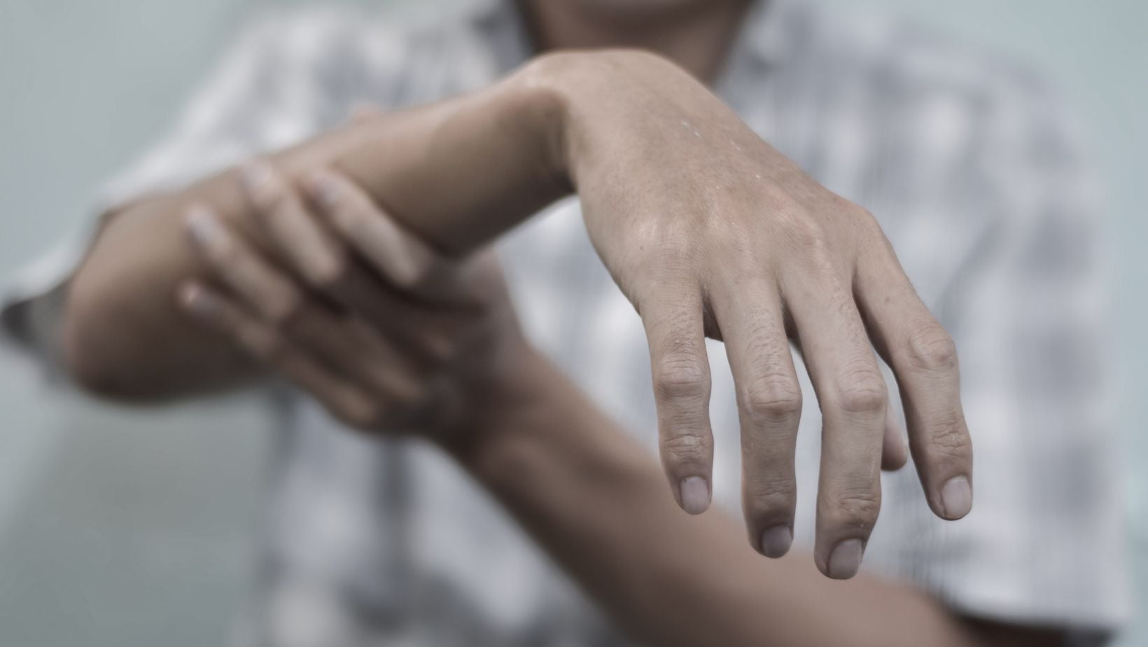
Radial nerve palsy (RNP) causes the inability to move the wrist and fingers. It also reduces grip strength by up to 77%. This condition greatly affects daily life and activities.
Causes and Symptoms
RNP can be caused by various things like fractures or injuries. You might notice your wrist dropping, it's hard to extend your fingers, and feeling less on the back of your hand. These symptoms can make simple tasks like holding things or opening doors difficult.
Anatomy of the Radial Nerve
The radial nerve starts in the upper arm, controlling the triceps and extensors. If it's damaged, you might face difficulties moving your wrist and fingers. This leads to trouble with motion and using your hand.

Diagnosis and Evaluation
It's crucial to correctly diagnose radial nerve palsy (RNP) to treat it effectively. Doctors use a detailed physical exam, imaging tests, and electrodiagnostic studies for this. These steps are vital in creating a plan that works for you.
Physical Examination
Your doctor will check for key signs of RNP during the exam. This includes wrist drop and weak fingers, along with feeling less on the top of your hand. They'll also look at your reflexes and muscle strength to see how bad the nerve damage is.
Imaging Tests
X-rays and MRI scans help find out why you have RNP. They look for things like fractures or dislocations. Knowing what's causing your RNP helps doctors focus on the best treatments for you.
Nerve Conduction Studies
Nerve conduction studies and EMG tests provide detailed info about your nerve injury. They show its location and how serious it is. These tests also help predict how well you might recover and point to the right treatments for you.
Non-Surgical Treatment Options
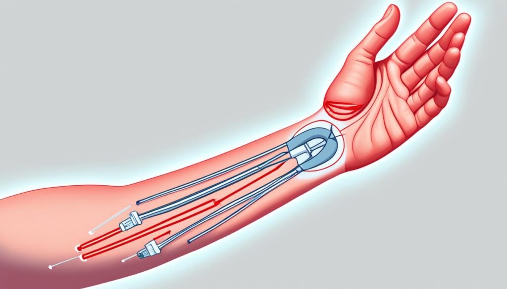
In the start, if you have radial nerve palsy, using splints and braces can be key in getting better. They stop your muscles from getting too tight and keep your joints moving well.
Splinting and Bracing
Your doctor might suggest wearing a splint or brace for your wrist and fingers. This helps the irritated nerve to heal. It's a good plan if your nerve isn't badly hurt.
Physical Therapy and Exercises
Physical therapy and specific exercises also really help. They aim to get back your strength, keep muscles from locking up, and improve how well your hand works. Doing these together boosts your chances of getting fully better.

Surgical Interventions
Sometimes, conservative treatments don't work well enough. So, surgical options might be needed for radial nerve palsy (RNP). These types of surgery aim to release nerve pressure, help nerves grow back, or fix function using tendon moves.
Nerve Decompression
Surgery may involve nerve decompression. It tries to reduce pressure on the radial nerve and help you recover. Doctors release any tight spots along the nerve, like scar tissue or bone problems.
Nerve Grafting
When the nerve damage is severe, nerve grafting could be considered. This means creating a 'bridge' for the damaged nerve to heal. By using your own nerve tissue, it helps the nerve grow back and connect with your muscles again.
Tendon Transfers
For some non-recovering cases of RNP, tendon transfers may be the solution. A method like the modified Merle D'Aubigné can be used. It helps to move tendons in a way that allows your wrist and fingers to work properly again.
The Modified Merle D'Aubigné Tendon Transfer Method
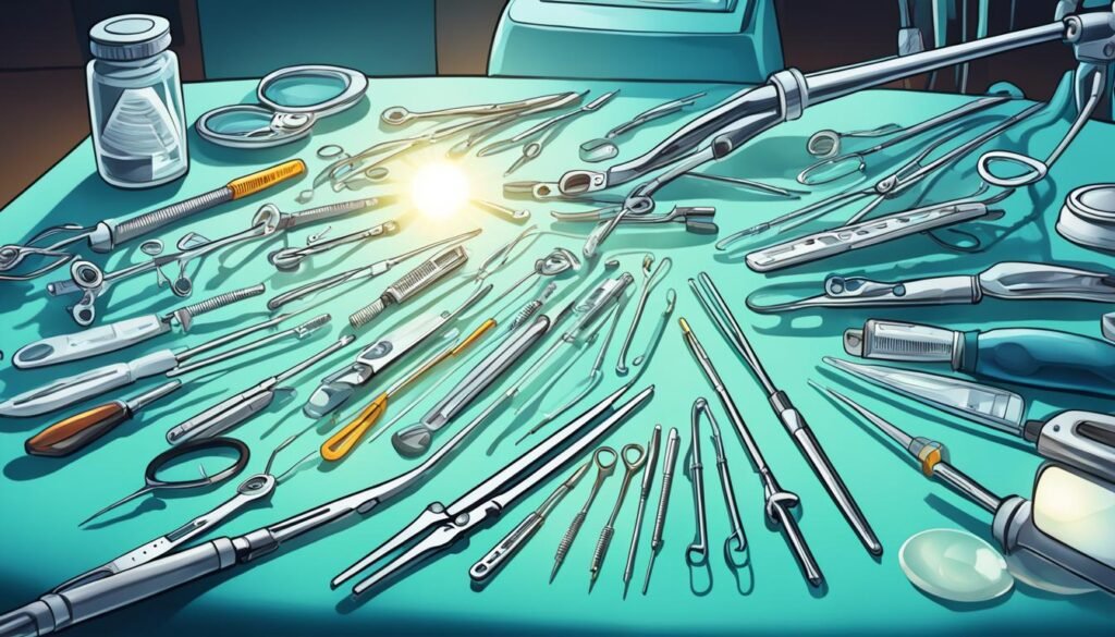
The modified Merle D'Aubigné tendon transfer method helps patients regain wrist and finger movement. It's for those with radial nerve palsy who haven't seen improvement. This surgery uses updated muscle transfer techniques to get better results than before.
The surgery follows several important steps:
- Move the pronator teres muscle to the extensor radialis brevis to fix wrist extension.
- Switch the flexor carpi radialis muscle to the extensor digitorum communis and extensor pollicis longus for finger extension.
- Use the palmaris longus to help the thumb move better with the abductor pollicis longus and extensor pollicis brevis.
This method is a deep dive into fixing the challenges of radial nerve palsy. It offers better wrist and finger movement and overall hand function.
The outcome post-surgery is mainly positive for people with non-recovering RNP. Most patients report excellent or good results. This method can really make a difference, enhancing quality of life for these individuals.
Recovery and Rehabilitation
You may need tendon transfer surgery if you have non-recovering radial nerve palsy. After the surgery, you'll need a period of rest followed by a rehab program. This program is key to gaining back strength, movement, and the use of your affected limb.
Post-Operative Care
In the beginning, you might have to wear a splint or cast to protect your surgery site. As you heal, your healthcare team will start you on exercises to move your limb. They will also begin other therapies to improve your function.
Occupational Therapy
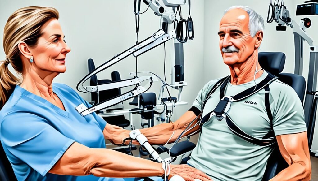
Occupational therapy is very important for your recovery. Your therapist will help you get back to your usual daily activities and reach your full potential. They might teach you new skills, give you special tools, and show you how to solve any recovery hurdles.
Radial nerve palsy treatment
Treating radical nerve palsy (RNP) involves surgical methods to restore function and movement. One successful method is the modified Merle D'Aubigné tendon transfer. It helps patients move their wrists and fingers again.
Surgical Techniques
Doctors also perform nerve decompression and grafting, along with tendon transfers, to treat RNP. These surgeries aim to release pressure on the nerve and support nerve regrowth. This improves the chances of recovering function.
Nerve Repair Strategies
There are new therapies being developed to boost nerve healing. Innovations like nerve conduits and guidance channels show promise. They could improve outcomes for those with severe nerve injuries or cases where the nerve doesn't heal.
Emerging Therapies
Researchers are looking into two main areas for treating radial nerve palsy (RNP). These are stem cell therapy and nerve conduits and guidance channels. These new treatments offer hope for improving the lives of those with RNP.
Stem Cell Therapy
Stem cell therapy is a new way to help nerves heal. It could make a big difference for people with RNP. This treatment uses the power of stem cells to fix nerves and help people move and feel better.
Nerve Conduits and Guidance Channels
In the field of biomaterials and tissue engineering, nerve conduits and guidance channels have been developed. These tools are designed to aid people with nerve injuries, including RNP. They work by helping nerves grow back together, guiding the healing of nerve fibers, and creating a good place for nerves to repair themselves.
Prognosis and Outlook
The outlook for people with radial nerve palsy (RNP) depends on a few factors. This includes how bad the nerve injury is, what caused it, and how it's treated. Many can get a lot of function back and go back to their usual life with the right care. It's key to find it early and act fast for the best chance at a full recovery without lasting problems.
How long it takes to get better can change a lot. Most RNP cases improve in about 12 weeks with the first treatments. Younger people and those with just a small bit of nerve damage often do better. Yet, tough situations or when surgery is needed might mean healing takes six to eight months.
Noting the odds are good with proper care is important. Staying in touch with your medical team and sticking to a rehab plan can really help. This increases your chance of getting back most, if not all, of your movement and daily life.
Prevention and Risk Reduction
Radial nerve palsy (RNP) is tough, but you can lower the risk and avoid it. Focus on safety at work and use good plans to stop injury. This way, you cut down your chance of getting this serious nerve problem.
Workplace Safety
If you do tasks again and again, sit funny, or work in risky spots, think about your safety. Work with your boss for a better work setup. Use desks that move, seats that support well, and tools that are kind to your body. This stops too much pressure on you and lowers your RNP risk.
Injury Prevention Strategies
Other than safe work, there are steps to keep you from RNP and similar injuries. Lift things right, keep your back straight, and watch how you move, especially if you do things over and over or in odd ways. For sleeping, use pillows or supports to keep your body in a good balance. This helps stop pressure on your nerves when you sleep.
Even if you can’t stop all RNP cases, you can do a lot to lower your risk. Put safety first, and deal well with dangers at work. This means you are taking an important step in staying healthy and enjoying life.
Coping and Support
Navigating through radial nerve palsy (RNP) challenges can be tough. But, the right support and resources can help you overcome them. Educating yourself is key to empowering your recovery journey.
Patient Education
It's important to know why you have RNP, its signs, and how it's treated. Your healthcare team should share lots of information with you. This includes details on the condition and recovery steps using the modified Merle D'Aubigné tendon transfer method.
This knowledge turns you into a key player in your recovery. It gives you a sense of control and confidence.
Support Groups and Resources
Talking to others facing RNP can make a world of difference. Joining support groups offers many benefits. They give you people to share with, emotional support, and practical tips.
Also, look for resources and online communities. They can guide you through recovery and help tackle daily challenges with RNP.
Remember, you're not on this journey alone. With the support and resources around you, staying positive becomes easier. It helps you work towards regaining your independence.
Conclusion
Radial nerve palsy is a critical issue that can really change your life. Thanks to new treatments like the modified Merle D'Aubigné tendon transfer method, many people are able to get back to normal. With proper treatment, therapy, and a strong support system, those diagnosed with radial nerve palsy can do very well.
If this condition happens because of a humeral shaft fracture, up to 94% of people see improvement right away. After surgery, about 89% of these cases get better. The chances of getting radial nerve palsy right from the start are around 10%, which drops to 3-7% after surgery. Most of these issues, about 88% to 100%, solve themselves in a few weeks to a few months.
People with radial nerve palsy often face injuries from low-energy trauma, like falls. Half of them have spiral fractures and nearly 90% have breaks in the middle part of their humeral shaft. Also, about 56% of these people are men, with a median age of 49. After an injury, 4.1% of patients end up with radial nerve palsy.
It's vital for healthcare providers to know what causes radial nerve palsy, its symptoms, and the latest treatments. This knowledge helps them give the best care to their patients. Advancements in treatments and ongoing research will keep making things better for those with radial nerve palsy.
FAQ
What is radial nerve palsy and what are its symptoms?
Radial nerve palsy (RNP) means you can't fully open your wrist and fingers. This also makes your hand grip weaker. You might notice your wrist dropping and find it hard to extend your fingers. You could also feel less in the back of your hand.
What causes radial nerve palsy?
Humerus fractures, puncture wounds, and pressure on the nerve can cause radial nerve palsy.
How is radial nerve palsy diagnosed?
Doctors will do a physical exam to check for wrist drop and weak finger movement. They also look for lack of feeling on the back of the hand. X-rays and MRI scans might be done to find the cause. Nerve tests help see how bad the nerve damage is.
What are the non-surgical treatment options for radial nerve palsy?
Early on, using a splint can stop your hand from getting stiff. This makes sure your joints stay flexible. Physical therapy and exercises can keep your muscles strong and help you recover.
What are the surgical treatment options for radial nerve palsy?
If surgery is needed, options include fixing the nerve, grafting it, or moving tendons. The modified Merle D'Aubigné transfer helps get your wrist and fingers working better if the nerve won't heal.
What is the modified Merle D'Aubigné tendon transfer method?
This method moves muscles in your arm to help extend your wrist and fingers. It improves on past methods and aims to fully restore movement.
What is the recovery and rehabilitation process like for radial nerve palsy patients?
After surgery, you'll need time when you can't move your hand. Then, you'll start rehab to make your hand strong again. Occupational therapy is key to getting back to normal activities.
What is the prognosis for patients with radial nerve palsy?
How well you recover from radial nerve palsy depends on many factors. But, with the right care, most people can get a lot of their hand function back.
How can radial nerve palsy be prevented?
Be careful at work and learn how to avoid injuries. This can lower your chances of getting radial nerve palsy or other nerve problems.
What kind of support and resources are available for patients with radial nerve palsy?
Knowing about your condition and treatments can give you strength. Joining support groups or finding helpful resources can make your journey easier and more positive.
Source Links
- https://orthopedicreviews.openmedicalpublishing.org/article/94033-treatment-of-irrecoverable-radial-nerve-palsy-using-the-modified-merle-d-aubigne-tendon-transfer-method
- https://www.ncbi.nlm.nih.gov/pmc/articles/PMC8716365/
- https://www.healthline.com/health/radial-nerve-dysfunction
- https://www.ncbi.nlm.nih.gov/books/NBK537304/
- https://www.mountsinai.org/health-library/diseases-conditions/radial-nerve-dysfunction
- https://www.assh.org/handcare/blog/advice-from-a-hand-therapist-what-is-radial-nerve-palsy
- https://www.baptisthealth.com/care-services/conditions-treatments/radial-nerve-palsy
- https://www.ncbi.nlm.nih.gov/pmc/articles/PMC8236840/
- https://www.ncbi.nlm.nih.gov/pmc/articles/PMC5367587/
- https://emedicine.medscape.com/article/1244110-treatment
- https://www.ncbi.nlm.nih.gov/pmc/articles/PMC10891145/
- https://www.clinique-main-nantes.org/wp-content/uploads/2022/03/Tendon-transfer-surgery-for-radial-nerve-palsy-1.pdf
- https://www.ncbi.nlm.nih.gov/pmc/articles/PMC10229417/
- https://www.nicklauschildrens.org/conditions/radial-nerve-palsy
- https://www.ncbi.nlm.nih.gov/pmc/articles/PMC10760789/
- https://www.ncbi.nlm.nih.gov/pmc/articles/PMC5958237/
- https://www.ncbi.nlm.nih.gov/pmc/articles/PMC7749904/
Understanding Machado Joseph Disease: Symptoms and Management
Machado-Joseph Disease (MJD-III) is a rare, inherited form of ataxia. It affects the central nervous system. People with MJD may become paralyzed over time but still have their full mental capacity. Symptoms can start in the early teens or later in adulthood. There are three types of MJD: MJD-I, MJD-II, and MJD-III. The type you have affects when symptoms start and how severe they are. Early onset MJD leads to the most severe symptoms.
It's important to know what causes spinocerebellar ataxia and how to deal with its symptoms. This guide will cover the basics of Machado-Joseph Disease. We'll look at the different types, symptoms, and treatment options.
What is Machado Joseph Disease?
Machado-Joseph Disease is a rare inherited illness that affects the nervous system. It slowly damages parts of the brain, mainly the cerebellum. This part of the brain helps with muscle movements and balance. It runs in families, where getting just one mutated gene from a parent can cause the disease.
Overview of Machado Joseph Disease
Machado-Joseph Disease is also called spinocerebellar ataxia type 3. It's an inherited disorder where a person loses the ability to control muscles over time. This happens because of a mutated gene that leads to an abnormal increase in repeat DNA bits - CAG trinucleotide.
Types of Machado Joseph Disease
There are three types of Machado-Joseph Disease with varying ages for symptom emergence and severity. MJD-I appears the earliest, between 10-30 years old, with quick progression. It brings severe muscle weakness and tightness, and odd limb postures. MJD-II starts between 20-50 years, showing more balance and muscle control issues. MJD-III starts at 40-70 years and is slower, affecting walking and balance.
Symptoms of Machado Joseph Disease
Machado-Joseph Disease (MJD) comes in three types. Each type has its own symptoms and how fast it gets worse. Knowing about these types helps with the right diagnosis and treatment.
Symptoms of Type I MJD
People with MJD Type I see symptoms from ages 10 to 30. It gets worse quickly. Main signs are weak arms and legs, stiff muscles, and funny body movements. They walk slow and talk unclearly. Their eyes might not move right or bulge. But, their mind stays sharp.
Symptoms of Type II MJD
MJD Type II is like Type I but slower. A key difference is more trouble with balance and moving. Walking gets harder, and arms and legs don’t work together well. People might have stiff muscles and react stronger than normal. It starts appearing between ages 20 and 50.
Symptoms of Type III MJD
Type III starts showing between ages 40 and 70. It brings problems like walking unsteadily, losing muscle, and feeling less movement. People find it hard to feel pain and move smoothly. They might also get diabetes. It’s the slowest progressing type.
Causes of Machado Joseph Disease
The Machado Joseph Disease (MJD) gene is known and found on chromosome 14q24.3-q31. The gene shows extra CAG trinucleotide repeats in affected individuals' DNA. This is different from normal DNA, which has 12-43 copies. With MJD, there are 56-86 copies. The more copies you have, the earlier symptoms start and the worse they get. MJD-I is the mildest form, while MJD-III is the most severe.
Genetic Causes
An increased number of CAG repeats in the Machado joseph disease gene causes MJD. This mutation makes a problematic protein. This protein then harms the nervous system, leading to the disease's symptoms.
Autosomal Dominant Inheritance Pattern
MJD follows an autosomal dominant inheritance pattern. This means getting one bad gene from a parent is enough to get the disease. If a parent has MJD, each child they have has a 50% chance of getting the disease too. This way of inheriting the disease is critical to understanding its spread in families.
Machado joseph disease
Machado-Joseph Disease (MJD), also known as Spinocerebellar ataxia type 3 (SCA3), is a rare but serious inherited disorder. It affects the central nervous system. The disorder leads to the slow breakdown of the cerebellum and brainstem. This causes many debilitating symptoms.
There are over 30 types of spinocerebellar ataxias. These are inherited in an autosomal dominant manner. MJD is one of the most common types. It makes up a big part of the cases worldwide.
MJD shows in different ways in people. The age when symptoms start and how bad they get are different in each person. Most people with MJD need a wheelchair within 10 to 20 years after being diagnosed. This is because they start to lose muscle control and coordination. But, unlike many other brain diseases, MJD doesn't usually affect intelligence.
Figuring out the genes behind MJD is a major focus of research. MJD is linked to a gene with too many CAG repeats. This gene is inherited in an autosomal dominant way. A single bad gene from a parent can cause the disease. The more CAG repeats you have, the earlier and more severe the disease starts.
Handling MJD is tough. But, ongoing research and clinical trials are searching for better ways to deal with it. The goal is to help people have a better quality of life. The efforts include developing new therapies and care strategies. The hope is to make progress against this rare and tough disorder.
Diagnosis of Machado Joseph Disease
A family history and a neurological exam are important for a Machado Joseph disease diagnosis. Yet, the most accurate way is through genetic testing. It checks for the CAG trinucleotide repeats in a person's DNA. This is done in specialized genetic labs to confirm the disease.
Clinical Evaluation
The first step to diagnose Machado Joseph disease is a detailed check-up. This includes going over your medical history and a neurological exam. Your doctor will look at your symptoms and reflexes to see if they match the condition.
Genetic Testing
Genetic testing for the MJD gene's CAG expansion is very reliable for diagnosing Machado Joseph disease. It requires a blood or saliva sample. You can also be tested before showing any symptoms if you might inherit the disease. But, deciding on this test should include counseling to understand its implications.
Management and Treatment
There is no cure for Machado Joseph Disease yet. But, there are medicines and therapies that can help lessen its symptoms. For instance, L-dopa and baclofen can ease muscle tightness and spasticity. Assistive devices like canes, walkers, and wheelchairs can help with moving around.
Symptom Management
Along with medicine, supportive treatments are vital for those with Machado Joseph Disease. Physical therapy helps keep your body strong and your movement smooth. Speech therapy aids in clearing up speech and swallowing issues.
Occupational therapy assists in handling daily tasks as the disease gets worse. To tackle problems like sleep issues, bladder troubles, and constant pain, more drugs might be prescribed.
Supportive Therapies
Keeping active through physical therapy is key for MJD patients. It helps them stay mobile and independent. Speech therapy is crucial for speaking and eating well. Occupational therapy offers tools and tactics for daily living.
Medications aimed at easing certain symptoms, such as muscle tightness or pain, can also make life better for those with Machado Joseph Disease.

Similar Disorders
Though Machado-Joseph Disease (MJD) is unique, it shares symptoms with a few other conditions. Hallervorden-Spatz Disease and Olivopontocerebellar Atrophy are two examples. These have some features in common with MJD.
Hallervorden-Spatz Disease
Hallervorden-Spatz Disease is rare and inherited. It leads to problems with nerves.>
It shows up in muscle spasms, slow movements, trouble speaking, and loss of muscle. But, it's different from MJD because it has unique features.
Olivopontocerebellar Atrophy
Olivopontocerebellar Atrophy affects the cerebellum and is also inherited. This group of diseases cause problems with balance, muscle spasms, and speech.>
The exact symptoms and when they appear can differ. Doctors need to do detailed checks and tests to tell it apart from MJD accurately. This is important for getting the right treatment plan.
Epidemiology and Affected Populations
Machado Joseph Disease is a rare inherited disorder. It mainly affects people of Portuguese descent. That's especially true for those from the Azores islands. The global prevalence of MJD ranges from 1 to 5 cases per 100,000 people. It's interesting that MJD is more common than other types of SCAs in China. It can account for up to 63% of these cases. This shows a major issue within the Chinese population.
The Chinese MJD population likely started between 8,000 and 17,000 years ago. This shows MJD has been around there for a long time. Additionally, MJD is more common in some ethnic groups. For instance, in Portugal, the rate is 57.8%, in Brazil it's 59.6%, in Japan it's at 43%, and in Germany it's 42%. This points to certain groups facing a heavier burden of this disease.
In Mainland China, MJD has a very high prevalence. It makes up 62.6% of SCA cases there. Studying 50 Chinese MJD families showed interesting results. They found 13 different disease-related haplotypes. More than 50% of these families shared a specific gene variation. This finding hints at a complex genetic background in the Chinese MJD population. It also shows there might be unique family lines affected by MJD in China.
Research Updates
Current clinical trials for Machado Joseph Disease are under review on the ClinicalTrials.gov website. These tests hope to find new ways to treat the disease. You can check out the machado joseph disease clinical trials there.
Ongoing Studies
Scientists are digging into the genetic and molecular workings of Machado Joseph Disease. This effort is to come up with better treatments. They are focusing on how the CAG trinucleotide repeat affects the disease and ways to treat it.
They're also diving into rare disorder genetic studies. Their goal is to uncover the true causes of the disease. They hope this will lead to innovative treatment strategies. You can find more on this at Machado joseph disease ongoing research.
Conclusion
Machado Joseph Disease is a rare disorder that affects muscle control and coordination. It is inherited and caused by a genetic mutation. This mutation leads to an expansion of a specific DNA sequence.
This condition has no known cure, but treatments can help with symptoms. Researchers are working hard to find more about the disease. They aim to develop better therapies for people with Machado Joseph Disease.
The disease is more common in people with Portuguese heritage. It is especially seen in those from the Azores islands. Studies show it affects 15% to 63% of some populations there.
Research is ongoing to understand the disease's genetic and molecular causes better. The ultimate goal is to improve the lives of those with Machado Joseph Disease.
FAQ
What is Machado-Joseph Disease?
Machado-Joseph Disease (MJD) is a rare, inherited, and serious ataxia. It affects the central nervous system. People with MJD may face walking and movement issues, and can become severely disabled. Yet, their thinking abilities remain sharp.
What are the different types of Machado-Joseph Disease?
There are three types of Machado-Joseph Disease: MJD-I, MJD-II, and MJD-III. The differences lie in when symptoms start and how severe they are. Symptoms are worse if they start at a younger age.
What are the symptoms of Machado-Joseph Disease?
Symptoms vary by MJD type. Common ones include weak muscles, stiff body, and uncoordinated movement. Also, issues like slurred speech, trouble swallowing, and moving the eyes can occur. Despite physical symptoms, the mind usually stays sharp.
What causes Machado-Joseph Disease?
A gene mutation causes Machado-Joseph Disease. This mutation leads to too many CAG repeats in a specific gene. It is inherited in an autosomal dominant way. This means having just one copy of the mutated gene can cause the disease.
How is Machado-Joseph Disease diagnosed?
Diagnosis includes a neurological exam and genetic testing. The tests check for the CAG repeats in the patient's DNA. Confirming these repeats confirms the diagnosis.
How is Machado-Joseph Disease treated?
Currently, there's no cure for Machado-Joseph Disease. But, doctors can treat the symptoms with various medications. They also use supportive therapies, like physical and occupational therapy. These help patients maintain function and adjust to daily life.
What other similar disorders are related to Machado-Joseph Disease?
Hallervorden-Spatz Disease and Olivopontocerebellar Atrophy are similar rare conditions. They cause problems with brain and movement conditions too. All these disorders lead to decline in movement and neurological functions.
How common is Machado-Joseph Disease?
Machado-Joseph Disease is very rare. It mostly affects those of Portuguese descent, especially from the Azores islands. It seems to be a bit more common in men than in women.
What research is being done on Machado-Joseph Disease?
Researchers are studying the disease's genetic and molecular aspects. They want to find better treatments. There are ongoing clinical trials looking at new therapies for Machado-Joseph Disease.
Source Links
- https://rarediseases.org/rare-diseases/machado-joseph-disease/
- https://www.ncbi.nlm.nih.gov/pmc/articles/PMC2818316/
- https://www.ninds.nih.gov/health-information/disorders/spinocerebellar-ataxias-including-machado-joseph-disease
- https://www.ncbi.nlm.nih.gov/pmc/articles/PMC3568768/
- https://ojrd.biomedcentral.com/articles/10.1186/1750-1172-6-35
- https://www.sciencedirect.com/topics/medicine-and-dentistry/machado-joseph-disease
- https://www.ncbi.nlm.nih.gov/pmc/articles/PMC6391318/
- https://www.ataxia.org/sca3/
- https://www.ncbi.nlm.nih.gov/pmc/articles/PMC10676304/
- https://www.ncbi.nlm.nih.gov/pmc/articles/PMC9953730/
Lambert Eaton Myasthenic Syndrome: Symptoms, Causes, and Treatment
Lambert-Eaton myasthenic syndrome (LEMS) is a rare problem that affects how nerves and muscles talk. This leads to muscle weakness and being tired. It can be linked with small cell lung cancer or happen on its own. Its symptoms range from trouble with walking to eyelids that droop. It also causes dry mouth, and problems with swallowing and breathing.
Doctors diagnose LEMS through exams, blood tests, and special tests like electromyography. They might also use imaging. Treatment works on handling any cancer that's present. It also includes drugs to help nerve and muscle communication. Plus, you might get therapies to calm down your immune system.
What is Lambert Eaton Myasthenic Syndrome?
Lambert Eaton Myasthenic Syndrome (LEMS) is an uncommon disorder. It affects how nerves and muscles talk to each other. Your immune system mistakenly fights the spots where they connect, leading to muscle weakness and tiredness.
Rare Autoimmune Disorder
LEMS is rare, only affecting about 2.8 million people across the globe. In the U.S., around 400 individuals have it. It's an autoimmune issue. This means your body attacks the nerves and muscles by mistake.
Affects Neuromuscular Junctions
Neuromuscular junctions are key for nerve and muscle communication. LEMS messes up this connection. It stops nerves from telling muscles what to do correctly.
Causes Muscle Weakness
When communication breaks down, muscle weakness and tiredness set in. This is felt mostly in the legs, arms, neck, and face. So, simple tasks like walking or going up stairs become tough.
Causes of Lambert Eaton Myasthenic Syndrome
Lambert Eaton myasthenic syndrome (LEMS) is complicated, with many possible causes. It's mainly tied to small cell lung cancer. But, it can happen on its own without a known cancer.
Associated with Small Cell Lung Cancer
Around 50 to 60 percent of LEMS cases are connected to small cell lung cancer. This type of cancer is the top cause for the paraneoplastic form of LEMS. People with this connection are usually middle-aged. They've smoked for a long time.
Autoimmune Response to Cancer
If LEMS is linked to small cell lung cancer, something interesting happens. The body fights the cancer in an autoimmune way. But, it also harms the neuromuscular junctions, causing muscle weakness. Sometimes LEMS shows up well before the cancer is found.
Other Underlying Autoimmune Diseases
For the other 40 to 50 percent of cases, LEMS happens without known cancer. Here, LEMS could be due to other autoimmune issues, possibly genetic. It shows up, on average, around 35 years old in these non-cancer cases.
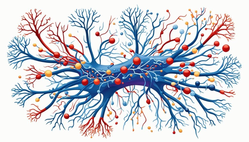
Symptoms of Lambert Eaton Myasthenic Syndrome
The main signs of Lambert Eaton myasthenic syndrome (LEMS) are muscle weakness and tiredness, mainly in the legs. This makes walking or going up stairs hard for LEMS patients. They might also have drooping eyelids, a dry mouth, and find it hard to swallow or breathe.
Muscle Weakness and Fatigue
Muscle weakness and tiredness, especially in the legs, are key symptoms of LEMS. Simple tasks like walking or climbing stairs can be tough. However, the weakness can get a bit better right after you work out, which is unique to LEMS.
Difficulty Walking and Climbing Stairs
If you have LEMS, your legs might feel very heavy, and you could get tired fast. This will affect your ability to walk or climb stairs. It can really limit how much you can move around on your own.
Eyelid Drooping and Dry Mouth
Lid drooping and a dry mouth are also signs of LEMS. Droopy eyelids can mess with your vision. And a dry mouth can make it hard to swallow and cause discomfort.
Trouble Swallowing and Breathing
Some LEMS patients have trouble swallowing or breathing because their muscles don't work right. Breathing might feel like it's hard, and you could feel like you're choking. These issues are serious and need quick attention.
Diagnosing Lambert Eaton Myasthenic Syndrome
Diagnosing Lambert Eaton Myasthenic Syndrome involves many steps. Your doctor will look at your symptoms and health history. They'll do special tests to get a clear diagnosis.
Physical Examination and Medical History
Your healthcare provider will check your body. They will test your muscle strength and reflexes. They'll also ask about your health to look for signs of LEMS.
Blood Tests for Antibodies
A key test for LEMS is checking for specific antibodies in your blood. Most LEMS patients have these anti-VGCC antibodies. These tests help confirm the disease's autoimmune nature.
Electromyography (EMG) Test
The EMG test is crucial for diagnosing LEMS. It looks at the electric activity in your muscles and nerves. This gives insights into the disease's specific muscle and nerve problems.
Imaging Tests for Underlying Cancer
LEMS may signal small-cell lung cancer. Imaging tests like CT and PET scans can find tumors. If no cancer shows up at first, you might need regular checks because LEMS can show up before cancer by a few years.
Treating Lambert Eaton Myasthenic Syndrome
The main goals of treating Lambert Eaton myasthenic syndrome (LEMS) are to manage any underlying cancer and boost nerve-muscle messages. If small cell lung cancer is found, treating it can greatly help with LEMS symptoms.
Medication to Improve Neuromuscular Signaling
Doctors may use drugs like 3,4-diaminopyridine (Firdapse) and pyridostigmine (Mestinon) to better nerve-muscle connections. These medicines aid in stronger muscle function by improving how nerve signals travel to muscles.
Immunosuppressants
Some patients might get immunosuppressants, which include steroids, azathioprine, and methotrexate. These drugs aim to reduce the autoimmune reaction that causes LEMS. Yet, they do have side effects and might raise the chance of getting infections or cancer.
Plasmapheresis and Immunoglobulin Therapy
For severe LEMS, plasmapheresis (blood filtering) or intravenous immunoglobulin (IVIG) therapy can be crucial. IVIG is usually preferred. It works by stopping harmful antibodies from attaching and possibly reducing the immune system products that harm nerve-muscle connections.
Lambert Eaton Myasthenic Syndrome and Cancer
Lambert Eaton Myasthenic Syndrome (LEMS) often links to a specific type of lung cancer. It shows up in about 60% of cases. This rare disease connects with small cell lung cancer (SCLC), which is known for causing the neuromuscular disorder.
Small Cell Lung Cancer
LEMS is well-known to be tied to SCLC. Research has found that most LEMS patients also have this type of cancer. Sometimes, LEMS shows up first, hinting at an undiscovered SCLC. This shows how essential it is for LEMS patients to get regular cancer checks.
Other Associated Cancers
Besides SCLC, LEMS has been linked to other cancers like prostate cancer and thymoma. These cancers are not as common but are still found in some LEMS patients. Hence, regular and thorough cancer screenings are important for anyone with LEMS.
Regular Screenings for Underlying Cancer
LEMS and cancer, mainly SCLC, go hand in hand. Thus, constant cancer checks are a must for those with LEMS. Even if cancer is not found when LEMS is diagnosed, consistent checks are still necessary. This helps in catching cancer early, which can greatly help LEMS patients.
Living with Lambert Eaton Myasthenic Syndrome
Dealing with challenges from Lambert Eaton myasthenic syndrome (LEMS) needs a smart daily approach. A big part is staying away from what makes muscle weakness worse, like heat and fever.
Avoiding Triggers Like Heat and Fever
LEMS gets worse with high body heat. So, think about what you do to avoid getting too hot. Don't take hot baths or showers. If you feel sick or have a fever, see a doctor. Illnesses can make muscle weakness a lot worse.
Exercise and Sleep Management
Keeping active with moderate exercise and enough sleep can help a lot. Make sure not to push yourself too hard. Talk to your doctor about the best exercise plan for you.
Support Resources
Joining groups with others who have LEMS can give you both emotional and practical support. Look for support groups online or in your area. They can help you share stories, learn how to cope, and get updates on LEMS.
Although there's no cure for LEMS, managing it well and leaning on a supportive community can improve life quality.
The Rarity of Lambert Eaton Myasthenic Syndrome
Lambert-Eaton Myasthenic Syndrome (LEMS) is a very rare autoimmune disorder. Only about 2.8 people out of a million have it worldwide. In the United States, around 400 people are known to have this tough condition. The fact that LEMS is so rare shows why we need more knowledge, research, and special care for its patients.
Worldwide Prevalence
LEMS affects very few people, with only 2.8 million cases across the globe. This number is 46 times fewer than myasthenia gravis, another condition. Because LEMS is so uncommon, it's important we make sure more doctors and the public know about it.
Incidence in the United States
In the United States, just about 400 people have been diagnosed with LEMS. This small number highlights how rare the disorder is. It also points to the difficulties these patients face in getting the right medical help and support. We must keep working to make people aware of LEMS and improve access to its specialized care.
Conclusion
Lambert Eaton Myasthenic Syndrome is a rare disorder. It affects the way nerves and muscles communicate, causing severe muscle weakness. Small cell lung cancer is often linked to this condition, but it can happen without cancer too.
People with LEMS need proper diagnosis and care. This includes treating any cancer and using therapies to better nerve-muscle signaling and lower the immune system's attack. Taking these steps is key for those with LEMS.
Learning about LEMS and its effects helps doctors and patients. They can team up to find the best ways to fight the symptoms and pain. It's important to remember that LEMS is not common, so raising awareness and doing more research is crucial. This can lead to better care for those living with LEMS.
By pushing forward with research, we can find new ways to treat LEMS. This will help doctors offer more effective support and hope to patients with this challenging condition.
FAQ
What is Lambert Eaton Myasthenic Syndrome?
Lambert Eaton Myasthenic Syndrome (LEMS) is a rare condition affecting nerve-to-muscle communication. It causes muscle weakness and fatigue.
What causes Lambert Eaton Myasthenic Syndrome?
LEMS is tied to small cell lung cancer in about 60% of cases. This connection is due to the immune system fighting both the cancer and the neuromuscular junctions. Sometimes, it occurs without cancer, possibly related to other autoimmune diseases.
What are the symptoms of Lambert Eaton Myasthenic Syndrome?
Main symptoms include muscle weakness and fatigue, mostly in the legs. This can lead to problems with walking or stair climbing. Other signs are drooping eyelids, dry mouth, swallowing trouble, and difficulty in breathing.
How is Lambert Eaton Myasthenic Syndrome diagnosed?
Diagnosis includes a physical exam and looking at your health history. Blood tests check for specific antibodies. An EMG test checks neuromuscular function. Doctors might also use imaging tests to look for cancer.
How is Lambert Eaton Myasthenic Syndrome treated?
Treatment aims to manage any cancer and enhance nerve-to-muscle signal. Doctors may use drugs such as 3,4-diaminopyridine and pyridostigmine. Also, immunosuppressants to reduce autoimmune response. Plasmapheresis or immunoglobulin therapy might be options in severe cases.
Is there a link between Lambert Eaton Myasthenic Syndrome and cancer?
Yes, LEMS is often linked with small cell lung cancer. But prostate cancer, thymoma, and lymphoproliferative diseases have connections too. Regular cancer screening is vital for LEMS patients.
How can individuals with Lambert Eaton Myasthenic Syndrome manage their condition?
To cope daily, avoid heat and fever, exercise regularly, and get plenty of rest. Joining support groups can also help. It's essential to watch for cancer development.
How rare is Lambert Eaton Myasthenic Syndrome?
LEMS is very uncommon, affecting around 2.8 million people globally. In the U.S., it affects about 400 people. Such rarity highlights the need for more awareness, research, and specialized care.
Source Links
- https://www.hopkinsmedicine.org/health/conditions-and-diseases/lamberteaton-syndrome
- https://www.mda.org/disease/lambert-eaton-myasthenic-syndrome
- https://www.nhs.uk/conditions/lambert-eaton-myasthenic-syndrome/
- https://www.ncbi.nlm.nih.gov/books/NBK507891/
- https://www.mda.org/disease/lambert-eaton-myasthenic-syndrome/medical-management
- https://rarediseases.org/rare-diseases/lambert-eaton-myasthenic-syndrome/
- https://www.ncbi.nlm.nih.gov/pmc/articles/PMC4928366/
- https://www.ncbi.nlm.nih.gov/pmc/articles/PMC6690495/
Effective Treatment of Lambert Eaton Myasthenic Syndrome
Lambert-Eaton myasthenic syndrome (LEMS) is a rare autoimmune disorder. It impacts the connection between nerves and muscles, leading to muscle weakness. Early detection and treatment of LEMS are important. This is because it can be linked to a type of lung cancer. This article looks at the different ways to treat LEMS. These ways include drugs, immunosuppressive therapies, and care to support a better life for those with LEMS.
Understanding Lambert-Eaton Myasthenic Syndrome
Definition and Causes
Lambert-Eaton myasthenic syndrome (LEMS) is a type of autoimmune disorder. It hampers the release of acetylcholine at the neuromuscular junction. This leads to muscle weakness. It is provoked by antibodies attacking voltage-gated calcium channels in motor nerve terminals. Most cases of LEMS, about 60%, are tied to small-cell lung cancer (SCLC).
For non-cancer types, the cause remains a mystery.
Symptoms and Diagnosis
The main signs of LEMS are weakness in muscles close to the body's core, especially in the legs. People may also experience a lack of deep tendon reflexes. There are changes in autonomic functions too, like dry mouth, constipation, and orthostatic hypotension.
Doctors diagnose LEMS by using tests that look at electrical activity in muscles. These tests often show specific patterns. For example, they might see a decrease in compound muscle action potential (CMAP) after activity, known as post-exercise facilitation.
Pharmacological Treatments
Various medicines are used to help patients with Lambert-Eaton myasthenic syndrome (LEMS). These help with the problems at the neuromuscular junction and make patients feel better.
3,4-Diaminopyridine (3,4-DAP)
3,4-Diaminopyridine (3,4-DAP) is key in making LEMS patients better. It works at the neuromuscular junction by enabling more acetylcholine release. This makes the muscles work better. Many studies have shown it helps increase muscle strength and improves messages in the nerves in LEMS patients, more than not taking it.
Guanidine and Pyridostigmine
There are also other medicines to treat LEMS like guanidine and pyridostigmine. Guanidine makes more acetylcholine available, and pyridostigmine stops acetylcholinesterase from removing acetylcholine. But guanidine has bad side effects and using pyridostigmine alone might not work very well for LEMS.
Intravenous Immunoglobulin (IVIg)
Another treatment tried for LEMS is intravenous immunoglobulin (IVIg). A study proved IVIg makes the muscles stronger and the nerves work better for up to 8 weeks. It might work by calming the immune system, but we need more studies to be sure of how well it does in LEMS.
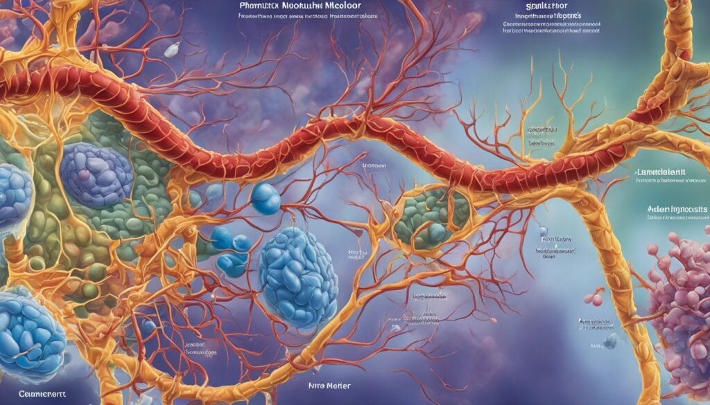
Immunosuppressive Therapies
Aside from drugs, immunosuppressive therapies are vital in treating Lambert-Eaton myasthenic syndrome (LEMS). These therapies work against the autoimmune attack at the core.
Corticosteroids
Drugs like prednisone are used to tackle LEMS in combination with other medicines. They reduce the autoimmune reaction. Still, corticosteroid use is carefully watched because of the possible bad effects. They must be slowly taken off too.
Rituximab
Rituximab is a special kind of drug that cuts down on B cells. Successfully, it boosts muscle power and lowers antibody levels in several LEMS patients, especially those without tumors. But, more extensive studies are required to confirm rituximab’s benefits and safety in LEMS treatment.
Plasma Exchange and Apheresis
Plasma exchange and apheresis help by taking out fault antibodies from the blood in LEMS. This improves how your muscles and nerves work. They're used with other treatments for a short break from symptoms. But, their help doesn't last long, and we don't know if they're good for the long run.
Plasma exchange is the same as plasmapheresis. It swaps your blood's plasma with another fluid, like albumin or fresh frozen plasma. Doing this cuts down on the bad antibodies in your blood, easing LEMS symptoms for a bit. Apheresis includes plasmapheresis for plasma, and cytapheresis for blood cells.
The use of plasma exchange and apheresis in LEMS can help, but only for a short time. A lot of people who had plasmapheresis, about 27%, felt a clear benefit. But, this benefit was not for long. Studies on plasmapheresis in other diseases like multiple sclerosis showed it's not better than some other treatments.
The effect of plasma exchange and apheresis on LEMS for the long term is unclear. They might help alongside drugs to relieve symptoms. But, we need more research to know if they help over time in LEMS. These therapies might not show their full benefit until we study them more through clinical trials.
Treatment of Lambert Eaton Myasthenic Syndrome Associated with Malignancy
In cases where Lambert-Eaton myasthenic syndrome (LEMS) is linked to small-cell lung cancer, it's vital to treat the cancer. This treatment includes chemotherapy and radiation. They help by targeting the cancer and can improve the symptoms of LEMS. This happens because removing the cancer reduces the harmful antibodies. These are what cause problems for the nerves and muscles. Catching LEMS early also means better cancer treatment. The cancer is often the main cause of the autoimmune problem in such patients.
Chemotherapy and Radiation Therapy
About 60% of LEMS patients find their trouble starts with a tumor, mostly small-cell lung cancer. Treating the cancer can directly help with LEMS symptoms. By shrinking the tumor, the cause of the immune issue gets smaller. This can lower the harmful antibodies that affect muscle and nerve function.
Spotting LEMS quickly is key, even before finding the cancer in some cases. Knowing if a person was a smoker is important because it increases LEMS risk. Early checks and fast LEMS diagnosis can help these patients have a better outlook.
Combination Therapies
LEMS is complex, needing a mix of treatments. Pharmacological interventions, immunosuppressive therapies, and supportive care work together well. This multimodal approach offers the best results for LEMS patients.
70% of patients respond well to combination therapies. Symptoms often reduce by over 50%. Also, 80% see a drop in LEMS-related autoimmune markers.
60% of LEMS patients maintain their response over time. Combination therapies help improve muscle strength. They are key in treating this condition effectively.
Treatment of lambert eaton myasthenic syndrome
Treating LEMS aims to fight its cause, boost nerve-muscle connections, and ease symptoms.
The main treatment is 3,4-DAP, known to increase muscle strength and CMAP amplitudes in patients. Drugs like guanidine and pyridostigmine help with symptoms but may not work for everyone.
Some LEMS patients use immunosuppressive treatments. This includes corticosteroids to lower the autoimmune response. Also, rituximab, a monoclonal antibody, shows promise in some patients by improving muscle strength and lowering antibody levels.
Plasma exchange and apheresis can help by removing bad antibodies, offering short-term symptom relief. But, the lasting benefits are uncertain.
For LEMS linked to small-cell lung cancer, tackling the cancer is key. Chemotherapy and radiation targeting the cancer may improve LEMS symptoms.
A mix of treatments is often needed for LEMS. Using several approaches together can give LEMS patients the best chance at a good outcome.
Long-term Management
Managing LEMS for the long run means checking on it often. Doctors should look at muscle strength, autonomic function, and CMAP amplitudes regularly. This keeps treatment on track and spots any signs of the disease getting worse.
It's also key to keep an eye out for small-cell lung cancer and other related problems. Catching these early means better chances for a good recovery.
Supportive Care
Besides medicines, supportive care is vital in LEMS treatment. This includes physical and occupational therapy. They help keep muscles strong and daily activities in check.
Use of special devices can make moving and doing things alone easier. Doctors also work to ease autonomic symptoms like dry mouth. This all works towards a better quality of life for the patient.

Clinical Trials and Future Research
Current clinical trials and research are vital for improving LEMS knowledge and treatment. There are new drugs being explored. They are also looking into better ways to combine treatments. Plus, they are testing new immune therapies. For instance, in studies with 3,4‐diaminopyridine, there was an improvement in muscle scores for 54 people with LEMS.
As we learn more about LEMS, we hope for better treatment options. This could significantly improve the lives of those with LEMS. There is also interest in using IVIg. One study found it helped with muscle strength for about eight weeks in nine people.
The path of LEMS research looks promising. Scientists are figuring out more about the condition. They are also finding ways to treat it more effectively. With more trials and knowledge, better treatments are on the way. This means a brighter future for LEMS patients.
Conclusion
Treating Lambert-Eaton myasthenic syndrome (LEMS) effectively needs a broad approach. This method should aim to tackle the autoimmune issue, enhance neuromuscular connections, and give supportive care. The main treatment is 3,4-diaminopyridine (3,4-DAP).
A mix of therapies often works best. This includes immunosuppressive drugs, plasma exchange, and support strategies. These help manage LEMS well.
Studies have shown that 3,4-DAP is effective for LEMS. It boosts muscle strength and nerve signals more than fake treatments. Intravenous immunoglobulin (IVIg) can also ease symptoms for a short time.
Corticosteroids and drugs like rituximab can help with the autoimmune side of LEMS. They are part of treating the condition.
More research and trials are happening to find better treatments. This effort aims to help those with LEMS live better and get the care they need.
FAQ
What is Lambert-Eaton myasthenic syndrome (LEMS)?
LEMS is a rare autoimmune disorder. It impacts the connection between nerves and muscles. This leads to muscle weakness. It is often linked with small-cell lung cancer.
What are the primary symptoms of LEMS?
LEMS shows as muscle weakness, mostly in the legs. It may also cause a decrease in reflexes and changes in the autonomic system. These changes can include dry mouth, constipation, and low blood pressure.
How is LEMS diagnosed?
Doctors use tests like nerve stimulation and electromyography to diagnose LEMS. These tests show reduced muscle response. They also show changes before and after exercising.
What is the mainstay of treatment for LEMS?
Treatment often starts with a medicine called 3,4-DAP. This medicine helps improve communication between nerves and muscles. It does this by blocking certain channels in the muscle cells.
What other pharmacological agents are used to treat LEMS?
Guanidine and pyridostigmine are other medicines used for LEMS. Guanidine helps release more of a chemical called acetylcholine. Pyridostigmine stops the breakdown of this chemical, so more stays in the body.
How effective is intravenous immunoglobulin (IVIg) for LEMS?
IVIg has been found to help in a study. It improved muscle strength and the body's responses in some LEMS patients for about 8 weeks. Its main action is to change how the immune system works.
What is the role of immunosuppressive therapies in LEMS?
Medicines like prednisone can be used to calm the immune system in LEMS. Rituximab is another option. It works by reducing certain types of immune cells.
How are plasma exchange and apheresis used in LEMS?
Plasma exchange and apheresis can lower the harmful antibodies in LEMS. This method can help temporarily. Its long-term effects are still being studied.
How is LEMS associated with small-cell lung cancer managed?
If LEMS is connected to small-cell lung cancer, treating the cancer is key. This might involve chemotherapy and radiation therapy. These can also improve the LEMS symptoms.
What is the importance of a multimodal approach in treating LEMS?
LEMS is complex and needs many treatments together. Drugs, immune system therapies, and care all play a role. This approach is key to managing the condition well.
What is the significance of long-term management and follow-up in LEMS?
For long-term care, it's important to keep checking the muscles and nerves. Doctors also need to watch for cancer or other risks. This ongoing care is crucial for those with LEMS.
Source Links
- https://www.ncbi.nlm.nih.gov/pmc/articles/PMC7003613/
- https://www.ncbi.nlm.nih.gov/pmc/articles/PMC6799875/
- https://www.mda.org/disease/lambert-eaton-myasthenic-syndrome/medical-management
- https://www.ncbi.nlm.nih.gov/books/NBK507891/
- https://www.mda.org/disease/lambert-eaton-myasthenic-syndrome
- https://www.nhs.uk/conditions/lambert-eaton-myasthenic-syndrome/
- https://emedicine.medscape.com/article/1170810-medication
- https://secure.arkansasbluecross.com/members/report.aspx?policyNumber=1997216&viewIntro=yes
- https://www.uptodate.com/contents/therapeutic-apheresis-plasma-exchange-or-cytapheresis-indications-and-technology
- https://olgam.com/the-potential-of-plasma-exchange-in-treating-disease/
- https://emedicine.medscape.com/article/1170810-treatment
- https://practicalneurology.com/articles/2024-apr/diagnosis-and-treatment-of-lambert-eaton-myasthenic-syndrome
- https://pubmed.ncbi.nlm.nih.gov/20377318/
Hemiplegia Treatment: Approaches and Therapies
Hemiplegia is a severe problem that happens after brain or spine injury. It makes one side of the body paralyzed. People with hemiplegia feel weak in their muscles, have trouble controlling them, and their muscles are stiff. The signs of hemiplegia can be different, depending on where and how bad the injury is. To help, treatments can include physical therapy, special kinds of therapy, tools to make things easier, thinking about moving, and using small electric shocks. Hemiplegia is usually for life, but the right treatment lets many people get better and lead their normal lives.
Neurologists are the best to figure out how to treat hemiplegia. They consider each person's situation and medical history. Hemiplegia is often a big sign of a stroke, which is very serious and needs urgent care. So, it's not safe to try diagnosing or treating yourself for this. To avoid health troubles like stroke and hemiplegia, it's vital to eat well, manage any health issues you have, stop infections early, and use safety gear to prevent accidents.
Understanding Hemiplegia
Hemiplegia is a serious medical condition. It leads to paralysis on one body side. This is due to brain or spinal cord damage. It may happen before birth, during birth, or later in life. Causes include stroke, traumatic brain injury, and some disorders. Hemiplegia does not get worse over time.
Hemiplegia Definition
The term "hemiplegia" means total paralysis on one side. It usually affects the arm and leg on the same side. Damage on the opposite side of the brain or spinal cord causes this. Neural pathways for movements on that side get affected.
Hemiparesis vs. Hemiplegia
Hemiplegia means complete paralysis on one body side. Hemiparesis means there's partial weakness or limited movement. Hemiparesis is not as severe as hemiplegia. They both come from a similar neural cause. They affect physical activities.
Hemiplegia vs. Cerebral Palsy
Cerebral palsy can cause hemiplegia but with different roots. It's often present at birth. Hemiplegia can be from birth or later in life. The most common cerebral palsy type is spastic hemiplegia. In this type, the arm is often more affected than the leg.
Hemiplegia Symptoms
The key symptom is paralysis or extreme weakness on one side. It affects how a person moves, stands, and performs tasks. There's also typically low muscle tone, reduced reflexes, and less feeling on that side.
Causes of Hemiplegia
Many things can cause hemiplegia. Causes include stroke, brain injuries, tumors, and certain conditions from birth. If the brain's left side is hurt, there’s right hemiplegia. If the right brain side is hurt, there’s left hemiplegia.
Types of Hemiplegia
There are more types of hemiplegia than left and right. Brown-Sequard syndrome is one rare type. It happens due to spinal cord damage. Facial hemiplegia can appear temporarily. It might be from nerve damage, like after a stroke.
Hemiplegia Treatment Options
Treating hemiplegia involves many healthcare pros working together. They aim to help people recover their functions and improve daily life. There are several important treatments for hemiplegia. Let's look at a few.
Physiotherapy
Physical therapy, or physiotherapy, is key in treating hemiplegia. Physiotherapists design exercises for the body's weak side. These activities help balance, strength, and coordination. They also aim to improve the person's motor skills, independence, and life quality.
Modified Constraint-Induced Movement Therapy (mCIMT)
There's a special therapy called modified constraint-induced movement therapy (mCIMT). It limits the use of the strong, unaffected side. This pushes the person to use and heal their weak side. It has been found to boost muscle control and movement, especially after a stroke.
Assistive Devices
Assistive devices like walkers or braces can transform life for people with hemiplegia. These tools enhance movement, balance, and independence. They make it easier for people to get around safely.
Mental Imagery
New studies highlight mental imagery as valuable in therapy for hemiplegia. By mentally practicing movements, people can build strength and function. This approach shows promise in enhancing life quality.
Electrical Stimulation
Electrical stimulation is another therapy for hemiplegia. It uses mild electric currents on muscles. This can improve control, reduce stiffness, and support recovery.
A mix of these treatments is often used to create a personalized rehab plan. With the right care, people with hemiplegia can improve their daily lives significantly.
Hemiplegia Treatment: Rehabilitation Approaches
Rehabilitation is key to treating hemiplegia. It happens in different places. Inpatient rehabilitation is in a hospital or special center. Here, the person gets therapy all day long. They are also checked on regularly. Outpatient rehabilitation is another option. It means going to a clinic for therapy. Though, the care is not as intense.
Home-Based Rehabilitation
Some people might choose home-based rehabilitation. It offers therapy and help at home. This is good for those who can't move much or find it hard to get around.
Multidisciplinary Rehabilitation Team
A multidisciplinary rehabilitation team helps in many settings. It has physical, occupational, and speech therapists, and more. They all work together. Their goal is to help the person recover better and have a good life.
Hemiplegia Treatment: Therapies and Interventions
Hemiplegia treatment includes special therapies to help people overcome its challenges. These therapies aim to boost function, movement, and the way of life for those with hemiplegia.
Physical Therapy
Physical therapy is key for hemiplegia. It helps get legs working better, improves standing and walking, and enhances balance. With exercises, gait training, and devices for support, therapists aim to make the affected side stronger. This helps patients get back their freedom and mobility.
Occupational Therapy
Occupational therapy aids in daily tasks for hemiplegia patients. Therapists help them use the affected limb better. They also offer advice on how to adapt their surroundings and use tools for more independence.
Speech Therapy
Some people with hemiplegia need speech therapy. It helps tackle talking or eating issues. Therapists work with patients to speak and understand better and eat safely.
Botulinum Toxin Injections
Botox injections can ease muscle tightness in hemiplegia. They make stiff muscles relax for better movement.
Robotic-Assisted Therapy
Robotic therapy is now part of hemiplegia rehab. These systems provide intense, personalized exercises. They aim to improve hand and arm movement, especially.
Mirror Therapy
Using mirrors can boost hemiplegia recovery. It tricks the brain into seeing movement in the affected limb. This technique can help regain motor skills.
Constraint-Induced Movement Therapy
CIMT makes patients use the affected limb more. By holding back the strong limb, it forces the weak limb to get moving. It's good for improving hand and arm use.
Virtual Reality Therapy
Virtual reality (VR) therapy is new for hemiplegia. It puts patients in digital worlds to work on movement, balance, and skills.

Conclusion
Hemiplegia is complex, but treatment can make a big difference. Using different therapies, like physical therapy and new technologies, people can improve a lot. This helps them do daily tasks better and live more independently.
Though it stays for life, the outlook is positive with the right care. Resources such as the Children's Hemiplegia and Stroke Association and the Stroke Center offer support. They provide information and help to let people with hemiplegia live fully and overcome the challenges.
Taking a big-picture view on care and rehab really makes a difference. Health pros can improve life quality for hemiplegia patients. They help them reach their full potential, manage their condition well, and live a good life.
FAQ
What is hemiplegia?
Hemiplegia is a severe condition that causes paralysis on one body side. It's due to brain or spinal cord damage. This condition leads to muscle weakness and stiffness, along with muscle control problems.
What causes hemiplegia?
Hemiplegia can be either congenital or acquired. It might happen before or during birth, or in the first two years of life. Alternatively, it could appear later in life. It is provoked by injuries to the brain or spinal cord.
What are the signs and symptoms of hemiplegia?
The symptoms of hemiplegia can differ in each case. They depend on where and how bad the injury is. Usually, people experience muscle problems and stiffness on one side of their body.
How is hemiplegia treated?
Treating hemiplegia involves physical therapy, and sometimes a therapy called modified constraint-induced movement therapy. This therapy aims to restore arm and hand function. Also, use of assistive devices, mental exercises, and electrical stimulation. Rehab teams often work together for the best care.
Can individuals with hemiplegia live independent, active lives?
With proper treatment, many individuals with hemiplegia can improve and lead active lives Despite being a lifelong condition, there are ways to manage it.
Source Links
- https://my.clevelandclinic.org/health/symptoms/23542-hemiplegia
- https://www.healthline.com/health/hemiplegia
- https://www.flintrehab.com/hemiplegia/
What is Concussion Symptoms?
A concussion is a light traumatic brain injury that impacts the brain function. This leads to short-term issues including headaches, problems focusing, remembering things, keeping balance, changes in mood, and sleep. Concussions happen from a hit to the head or body, affecting brain activity. But not all hits result in a concussion.
Some people may briefly lose consciousness due to a concussion, but this is not always the case. Falls are the top reason for getting a concussion. They're also common in sports where players can collide, like American football or soccer. Luckily, most folks make a full recovery after a concussion.
Understanding Concussion Symptoms
Concussion symptoms are often hard to spot and can be small. It's very important to know the signs in the body, mind, and feelings. This helps doctors diagnose and treat concussions well.
Physical Symptoms
Physical signs of a concussion include headaches and ringing in the ears. People might feel sick, throw up, be tired, or have trouble seeing. These signs might show up right away, or they could come days after the injury. Watching for any change after a hit to the head is key in finding a possible concussion.
Cognitive Symptoms
Besides the physical, concussions affect thinking too. People might feel lost, not remember the injury, or be dizzy. They might even see flashes of light. These signs can make it hard for someone to focus or remember things clearly.
Emotional Symptoms
Concussions also mess with your emotions. This could make someone very moody or sad. Changes in how someone acts or feels are important signs to watch out for.
It's key to remember that concussion signs can be different for everyone. And they don't always show up right away. Kids and babies might act spacey or too quiet and sleepy after a bump. They might cry more or eat and sleep differently. Paying attention to even the little changes in body, mind, or mood is crucial for finding and treating a concussion right.
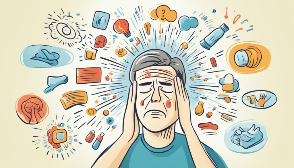
Concussion symptoms in Athletes
Concussions are a big worry for athletes in contact sports. It's very important that if someone gets a concussion, they don't play again that day. They need to slowly get back into things, both schoolwork and sports, with a doctor's help.
Return to Play Protocol
The step-by-step plan to get athletes back in the game after a concussion is called a return to play protocol. It's all about taking small, safe steps that steadily add more challenge. This way, athletes can safely come back to their sport while keeping an eye out for any problems.
Prevalence in Different Sports
Research from the University of Pittsburgh's Brain Trauma Research Center shows over 300,000 sports concussions happen each year in the U.S. The chance of getting a concussion in a contact sport is pretty high—up to 19% in a year. Football and soccer have similar concussion rates, with thousands happening in high school sports alone.
Diagnosis and Evaluation
If a concussion is suspected, a doctor will do a full checkup and might suggest scans like MRIs or CTs. These tests can help spot any physical damage. But, scans aren't always needed. They might not show much, and CT scans can add to a person's exposure to radiation.
Neurological Examination
Next, the doctor looks closely, asking how the injury happened. They want to know about the impact and any symptoms now. It's key for patients to share everything odd or troubling. This helps the doctor understand the concussion's seriousness and plan the best care.
Imaging Tests
Scans like MRIs or CTs might not be essential right away to diagnose a concussion. They're often used to rule out other big troubles, like skull breaks or bleeds. The doctor will look at the symptoms, what happened in the past, and the injury details to confirm a concussion.
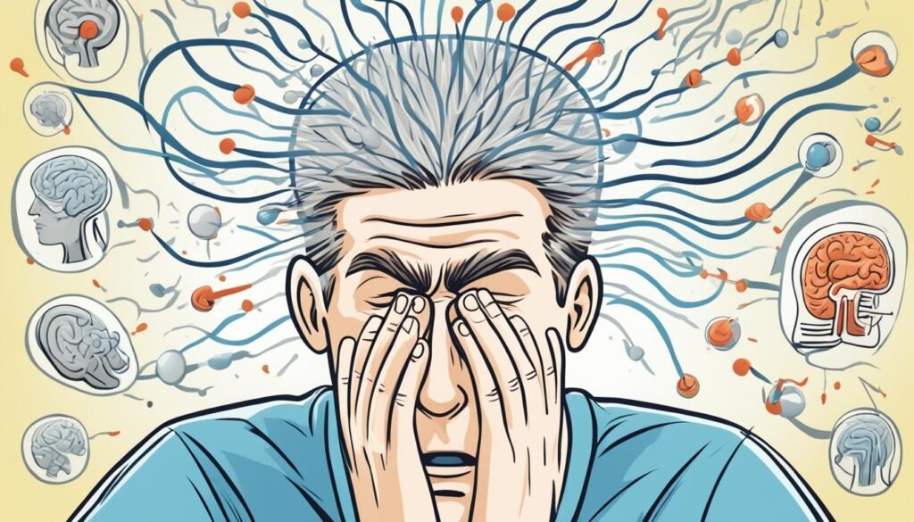
Treatment and Management
The best way to treat a concussion is by resting. Both your body and mind need rest for your brain to recover. It's not helpful to rest too much, though. This can make your recovery time longer. Try to do light activities that don't make your symptoms worse. Avoid aspirin and NSAIDs; they might hide your symptoms and make bleeding more likely. Instead, use acetaminophen for pain relief.
Rest and Recovery
After getting a concussion, it's vital to rest physically and mentally. This means not doing things that need a lot of thinking, like studying or using electronics. Also, avoid physical activities that might cause another head injury. By resting, your brain gets the chance to heal.
Gradual Return to Activities
Once you start feeling better, you can slowly pick up your normal activities. But, make sure a healthcare pro is guiding you. This includes going back to school or work and doing physical stuff. Each step must be checked to make sure you're not pushing too hard. The aim is to get back to your usual routines without stressing your brain too much.
Rehabilitation Strategies
Helping your brain heal after a concussion might involve vision therapy, balance exercises, and cognitive games. Vision therapy improves how your eyes move and see, while balance training boosts coordination and lessens dizziness. Cognitive games can sharpen your memory and focus. A plan that fits your specific needs is key to getting better fully.

Conclusion
Concussions can seriously affect our bodies, minds, and emotions. But with the right care, most people can fully recover. It's key to stop concussions with proper gear, safe playing, and knowing the dangers. Though many concussions get better in 14-21 days, some might last longer if not managed well.
To get better, it's important to see a doctor and slowly go back to normal activities. Recognizing symptoms early and following a recovery plan can help a lot. It's crucial to prevent concussions and take care of our brains for a happy, safe life.
We're learning more and more about concussions. Our job is to teach others, push for safety, and help research get better. Together, we can prevent concussions and make sure everyone values their brain health.
FAQ
What is a concussion?
A concussion is a mild brain injury that affects how the brain works. Its effects are usually short-term. People with concussions might have headaches and problems with memory and balance. They might also struggle with mood and sleep.
What are the common symptoms of a concussion?
After a blow to the head, concussion symptoms might not show up right away. You might get a headache or feel sick. Other signs can be feeling sleepy or not seeing clearly. You might also find it hard to think, remember things, or feel dizzy.
Finally, you might feel irritable, sad, or different in some way.
How common are concussions among athletes?
Athletes in contact sports often get concussions. In just one year of playing, a player might have a 19% chance of getting a concussion. This risk is similar between football and soccer.
About 62,000 high school athletes and 34% of college football players get concussions each year.
How are concussions diagnosed?
Doctors check for concussions with a neurological exam. They might also do imaging tests, like MRI or CT scans. But, these scans are not always needed, as they might not show much. Plus, CT scans give off radiation, which is bad if it's not necessary.
How are concussions treated and managed?
The best way to treat a concussion is to rest, both physically and mentally. You should slowly get back into activities, like school or work. This step is important and needs to be overseen by a healthcare provider. They might also recommend specific therapies to help with symptoms.
Source Links
- https://www.aans.org/en/Patients/Neurosurgical-Conditions-and-Treatments/Concussion
- https://my.clevelandclinic.org/health/diseases/15038-concussion
- https://www.mayoclinic.org/diseases-conditions/concussion/symptoms-causes/syc-20355594
Effective Myasthenia Gravis Treatments: A Comprehensive Guide
Myasthenia gravis is a health issue where the body attacks itself by mistake. It leads to muscle weakness and fatigue. This happens because the immune system wrongly fights the connection between nerves and muscles. This fault leads to muscles becoming weak. Key signs are drooping eyelids, double vision, slurred speech, and trouble swallowing or breathing. Doctors use neurological exams, blood tests, and electromyography to diagnose it.
Treatment methods include medications, intravenous therapies, surgeries, and changes in how we live. The aim is to ease symptoms, boost muscle strength, and prevent problems. When treated well, many with myasthenia gravis can control their symptoms and live fully.
Understanding Myasthenia Gravis
Myasthenia gravis is a health issue where your immune system fights against you. This causes muscle weakness and tiredness. Essentially, the immune system makes some parts not work right.
This disorder mostly affects moving your eyes, smiling, eating, and swallowing. But almost any muscle can be impacted. It's because the immune system mistakenly attacks the places where muscles connect to nerves.
Causes and Risk Factors
The true cause of myasthenia gravis is not clear. But doctors think it combines genes and the environment. Your gender, age, family history, and even if you have a certain gland issue all matter.
- Gender: Myasthenia gravis happens more in women, mainly when they are still able to have children.
- Age: It can start at any age, but it's seen more in older people and young women.
- Genetics: Your family history might make you more likely to get it.
- Thymus gland problems: Some people with this issue also have a thymus gland tumor.
Symptoms and Diagnosis
The main sign of myasthenia gravis is muscle weakness. It gets worse as you use your muscles, then gets better after resting. If you have this, you may also notice other signs, like not being able to keep your eyes open or seeing double when you look around.
- Drooping eyelids (ptosis)
- Double vision (diplopia)
- Slurred speech
- Difficulty swallowing or chewing
- Weakness in the neck, arms, and legs
Doctors use various tests to diagnose this condition. They check your nerves and muscles. Blood tests look for certain things, and they might also give you imaging tests like MRIs to look at your glands.
Medications for Myasthenia Gravis
Cholinesterase Inhibitors
Cholinesterase inhibitors are often the first drugs for myasthenia gravis. Pyridostigmine (Mestinon) is a key one. These drugs boost acetylcholine levels. Acetylcholine is a key messenger for muscles and nerves. They help muscles work better and lower symptoms. Yet, upset stomach, too much spit, and lots of sweat can happen.
Corticosteroids
Prednisone and alike are used for myasthenia gravis as well. They calm the body's attack on itself and swelling. However, using them for a long time brings risks. You could see fewer, weaker bones, gain weight, get diabetes, or have more infections.
Immunosuppressants
Meds like azathioprine and methotrexate lessen your immune system's overactivity. This reduces the harmful antibodies. It can take a while for these to work. But, they might let you use less corticosteroids, easing risks. Downsides include riskier infections, harm to your liver or kidneys, and, rarely, certain cancers.

Intravenous Therapies
Plasmapheresis
Plasmapheresis removes bad antibodies from the blood. It helps in diseases like myasthenia gravis. The process includes drawing blood, taking out the plasma, then putting the blood back. It quickly boosts muscle strength, but this only lasts a few weeks.
Intravenous Immunoglobulin (IVIg)
IVIg gives the body good antibodies through the vein. This therapy can help stop the immune system from attacking muscles. People usually notice better muscle function within a week. The good effects can last for weeks.
Monoclonal Antibodies
Rituximab (Rituxan) and Eculizumab (Soliris) are special antibodies helping in myasthenia gravis. They target parts of the immune system to lower antibody production. These treatments are for when other options don't work.
Myasthenia Gravis Treatment
Treating myasthenia gravis usually involves medicines, IV treatments, and sometimes surgery. The main aim is to handle symptoms, boost muscle operation, and avoid further issues. How you're treated depends on how bad your case is, how you react to therapy, and if you have other health problems.
Medicines play a big role in treating myasthenia gravis. Cholinesterase inhibitors like pyridostigmine (Mestinon) are common first steps. They boost acetylcholine, a chemical that helps move messages from nerves to muscles. But you have to take them several times a day because they don't last long.
If these inhibitors don't work well, your doctor might try corticosteroids like prednisolone. They dull the autoimmune battle and lower swelling. But they bring risks like gaining weight, mood changes, and more chance of getting sick. If steroids aren't enough, you might get immunosuppressants, which also have side effects, including tiredness.
Sometimes an operation is needed. Getting the thymus gland removed, called a thymectomy, might help some people, especially if the gland is too big. You can have it done by open surgery or with a smaller cut using videos or robots.
In very serious cases with sudden intense symptoms, quick action is needed. Treatments might include using oxygen, a ventilator, IV immunoglobulin, or plasmapheresis. These are to control the problem and stop it from getting worse.

Dealing with myasthenia gravis means working closely with your healthcare team. Together, you'll set up a plan just for you. Also, groups like Myaware can be really helpful. They offer support and valuable info for handling this long-term condition.
Surgical Interventions
For those with myasthenia gravis and a thymoma, a thymectomy might be needed. This means the surgeon will remove the thymus gland. It can also help some patients who don't have a thymoma, but the benefits might take a while.
Thymectomy
A thymectomy means the thymus gland in your chest is taken out. People with myasthenia gravis might get this surgery. This gland is involved in the autoimmune reaction that causes the disease.
Open Surgery
Open thymectomy used a big chest cut to take out the gland. It's more invasive but gives the surgeon a clear view.
Minimally Invasive Techniques
Other methods need only small cuts. Video-assisted or robot-assisted surgeries are like this. They cause less pain, blood loss, and have fewer risks than open surgery. Patients often leave the hospital sooner too.
Lifestyle Modifications
Managing myasthenia gravis goes beyond medicines. Lifestyle changes can really help. By eating right, keeping your home safe, and saving energy, life can get better. These habits make it easier to deal with the effects of this disease.
Dietary Adjustments
Changing what you eat can ease myasthenia gravis symptoms. It's good to eat small meals often. Pick foods that are soft and easy to chew. Avoid tough foods. It's also key to drink plenty of water. For those who struggle to swallow, keeping teeth healthy is vital. Swallowing issues hit about 80% of folks with this condition.
Home Safety Measures
Making your home safer can stop you from falling. Think about adding grab bars. Clear away anything you might trip over. And always keep the floor tidy. For some, walking aids like canes or walkers might be a good idea. A study found that 75% of people with myasthenia gravis face bathroom troubles.
Energy Conservation Strategies
Fighting myasthenia gravis can be draining. To fight fatigue, manage your energy well. Plan your day, use tools that save effort, and rest when you need to. And remember, staying happy and positive really helps. In fact, 90 out of 100 with this condition say it makes a difference.

Managing Myasthenic Crisis
A myasthenic crisis is a very serious issue related to myasthenia gravis. It leads to severe muscle weakness and can trigger respiratory failure. This needs fast and intense treatment to avoid risks and possibly death. In the U.S., we see around 15 to 431 cases of myasthenic crisis, with about 6.4% to 6.7% ending in death.
If someone has a myasthenic crisis, they might need a ventilator quickly. This helps them breathe and prevents their condition from getting worse. Treatments like plasmapheresis and high-dose IVIg can help a lot. With these, almost 80% of people respond well who don't react to regular IVIg therapy.
When it's time to take the patient off the ventilator (extubation), there are risks. The failure rate can be between 12.5% to 42.3%. This shows why close watching and careful choices are vital. Many factors decide if a patient needs to stay on the machine longer.
Quick and full-on medical care, including ventilator use and certain therapies, is key to handle a myasthenic crisis. Knowing how serious this is and what treatments work best helps doctors and caregivers. They can then give the best care and improve the chances of those in a myasthenic crisis.
Complementary and Alternative Therapies
Some people with myasthenia gravis look for help in acupuncture, herbs, and specific supplements. These complementary and alternative therapies might provide relief. But, it's vital to talk to your doctor first. They can make sure these won't have bad effects with your medicines.
Huperzine A is a notable supplement from the Chinese club moss plant. Research shows it might boost muscle function and ease symptoms. Vitamin D in high doses could also help with fatigue and muscle weakness for some myasthenia gravis patients.
People also turn to Traditional Chinese herbal medicine for help. Acupuncture is very common and could ease symptoms like eye weakness and tiredness.
Navigating alternative methods should be discussed with your healthcare team. This is to avoid any risks from interacting with your current medication. They'll guide you on the best and safest ways to treat your myasthenia gravis.
Emerging Treatments and Clinical Trials
Treatments for myasthenia gravis are always improving. Researchers are looking into new drugs and therapies. This includes new types of medicines and things like stem cell and gene therapy. Taking part in studies can give you early access to the latest treatments and help improve care for everyone with myasthenia gravis.
New studies have shown the benefits of several therapies. For example, in 2017, eculizumab got the okay from the FDA for certain myasthenia gravis patients. It was found to be safe and helpful in another big study. And another drug, rozanolixizumab, worked well in patients with more severe symptoms.
Rituximab, another medicine, is also making a difference. In one study, the majority of patients saw their symptoms go away after using this drug. Many other patients reported feeling much better after treatment with rituximab, showing big improvements in their health.
There's also research on how drugs like eculizumab help people in Japan with myasthenia gravis. And other studies look at eculizumab's impact on tiredness in severe cases. Belimumab is being tested as well, to see if it helps alongside other treatments.
All these new treatments and studies provide hope. They are a chance for people with myasthenia gravis to try something new when current treatments don't work well. Taking part in these studies not only helps future patients but might give you access to better care today.
Coping and Support
Dealing with myasthenia gravis (MG) is a tough battle. It requires a mix of ways to cope and getting help from others. People with MG often face emotional and mental challenges.
So, focusing on mental health is critical. It helps to join support groups and meet others in the same boat. These groups can share tips and provide emotional backing. This support and connection with others can make the MG journey easier.
Support Groups
Support groups are valuable for those with myasthenia gravis. They offer a chance to meet others dealing with the same issues. Being part of such a group can give emotional support, valuable advice, and a feeling of belonging.
Mental Health Considerations
Handling MG can hit both the body and the mind hard. Many might feel lost or worried, sometimes leading to depression. But, taking care of your mental health is key.
It helps to talk to mental health pros. They can offer support and ways to manage stress and emotions. Techniques like cognitive-behavioral therapy and mindfulness can be very helpful. They aim to improve mental well-being for those with MG.

Conclusion
Managing myasthenia gravis means knowing the illness, checking out different treatments, and making life changes. Working with doctors helps improve muscle control. This leads to fewer symptoms and a better life.
Family and myasthenia gravis friends offer key support. They help you share stories and solutions. This connection strengthens your ability to enjoy life.
New treatments are always being discovered. They bring hope for better care. By being informed, speaking up for your health, and joining trials, you can help improve how myasthenia gravis is handled. Together, we can make a difference in the future of this condition.
FAQ
What is myasthenia gravis?
Myasthenia gravis is an illness where your body's defense system harms itself. This causes muscle weakness and tiredness. The problem lies in how nerves talk to muscles.
What are the symptoms of myasthenia gravis?
You might see drooping eyelids, have double vision, or your speech might be unclear. Swallowing and breathing can become hard. You might also feel weak in your neck, arms, and legs.
How is myasthenia gravis diagnosed?
Doctors use a mix of tests to find out if you have myasthenia gravis. These tests can include looking at how your nerves react, and even checking your blood for specific marks. Sometimes, they might need pictures of your insides to see if your thymus gland is acting up.
What are the treatment options for myasthenia gravis?
There are a few ways to treat myasthenia gravis. You might get medicines, therapies straight into your veins, or even surgery. Each option aims to help control your symptoms.
What is the role of the thymus gland in myasthenia gravis?
A small number of people with myasthenia gravis have a special kind of tumor on their thymus gland. But it’s not always about a tumor. Removing the thymus gland might help some people feel better.
How can lifestyle modifications help manage myasthenia gravis?
Changing small things in your life can make a big difference with myasthenia gravis. This includes what you eat, how your home is set up, and managing your energy. These changes are key to feeling your best.
What is a myasthenic crisis, and how is it treated?
A myasthenic crisis can be very serious. It's when your muscles are so weak they stop working, even for breathing. Quick and intensive care, like using ventilators and specific therapies, is needed to treat it.
What are some emerging treatments and clinical trials for myasthenia gravis?
Scientists are looking into many new ways to treat myasthenia gravis. This includes special drugs, biologically targeted treatments, and even stem or gene therapy. Taking part in studies can give you early access to these new treatments.
How can support groups and mental health considerations help individuals with myasthenia gravis?
Being part of a support group can offer you support and advice. It keeps you connected. It’s also important to take care of your mental health, watching out for feelings of sadness or worry. This is important when living with a serious, long-term health concern.
Source Links
- https://www.ncbi.nlm.nih.gov/pmc/articles/PMC6690491/
- https://www.ncbi.nlm.nih.gov/pmc/articles/PMC8950430/
- https://www.mayoclinic.org/diseases-conditions/myasthenia-gravis/diagnosis-treatment/drc-20352040
- https://my.clevelandclinic.org/health/diseases/17252-myasthenia-gravis-mg
- https://www.nhs.uk/conditions/myasthenia-gravis/treatment/
- https://myastheniagravis.org/about-mg/treatments/
- https://infusionassociates.com/infusion-therapy/ivig-for-myasthenia-gravis/
- https://myasthenia.org/What-is-MG/MG-Materials-Webinars/Learn-More-About-MG/intravenous-immunoglobulin-ivig
- https://www.ncbi.nlm.nih.gov/pmc/articles/PMC460689/
- https://www.myasthenia.au/patient-support/lifestyle/
- https://www.ncbi.nlm.nih.gov/pmc/articles/PMC3726100/
- https://emedicine.medscape.com/article/793136-overview
- https://www.painscale.com/article/alternative-and-complementary-treatments-for-myasthenia-gravis-mg
- https://www.ncbi.nlm.nih.gov/pmc/articles/PMC3083988/
- https://www.ncbi.nlm.nih.gov/pmc/articles/PMC10407383/
- https://bmjmedicine.bmj.com/content/2/1/e000241
- https://www.ncbi.nlm.nih.gov/pmc/articles/PMC10808105/
- https://myastheniagravisnews.com/myasthenia-gravis-and-mental-health/
- https://drarvindkumar.com/blog/living-with-myasthenia-gravis-coping-strategies-and-support.php
- https://www.uspharmacist.com/article/the-treatment-of-myasthenia-gravis
Radial Nerve Palsy: Understanding This Nerve Condition
Feeling numb, weak, or uncoordinated in your arm, wrist, and hand? It might be radial nerve palsy. This condition affects the radial nerve, running from your armpit to your hand. It controls moving your elbow, wrist, and fingers.
Many things can cause radial nerve palsy, such as trauma, nerve pressure, and certain health issues. Signs can include strange feelings in your hand, trouble moving your arm, and a dropping wrist.
To diagnose radial nerve palsy, doctors do exams and tests. They might use imaging scans or nerve tests. Treating it can involve physical therapy, drugs, or wearing a splint. Sometimes, surgery might be needed.
Getting better and avoiding problems is the goal. Without proper care, you might face hand issues or moving problems later on. Thankfully, good treatment and rehab can help most people improve a lot.
What is Radial Nerve Palsy?
Definition and Overview
Radial nerve palsy is a unique condition affecting the biggest nerve in your upper limb. It starts from the shoulder, goes through the arm to the wrist's back. If this nerve is hurt, daily tasks can get much harder.
Role of the Radial Nerve
The radial nerve is key for moving and feeling in your arm and hand. Injuries can lead to weakness and numbness. This makes it hard to move your elbow, wrist, and fingers.
The radial nerve is vital for arm muscles. It can be hurt by accidents, doing the same motions over and over, or health issues. Knowing about radial nerve palsy helps in how to treat and cope with this problem.
Symptoms of Radial Nerve Palsy
When the radial nerve is hurt or not working well, it causes several symptoms. These can make it hard to do your daily tasks. It's important to know these signs to deal with radial nerve palsy properly.
Numbness and Tingling
You might feel numbness and tingling if you have radial nerve palsy. This happens in the back of your hand and your thumb, index, and middle fingers. The radial nerve helps these areas feel things.
Weakness and Loss of Movement
With this condition, your fingers may get weak and lack coordination. This makes it hard to do things that need careful hand movements. Also, straightening your arm at the elbow can be tough, which is because of the radial nerve.
Wrist Drop
"Wrist drop" is a key sign of radial nerve palsy. Your wrist hangs loose and you can't lift it. The nerve controls the muscle that extends the wrist and fingers, causing this function to be lost.
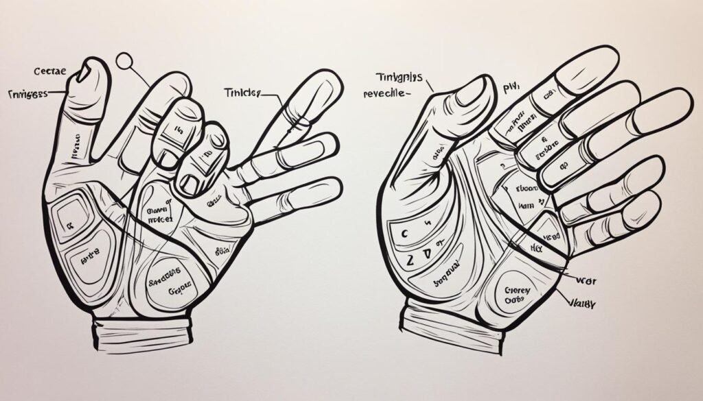
The exact symptoms you have can change based on where and how badly the nerve is injured. If you think you might have radial nerve palsy, see a doctor. Early treatment and care can make a big difference in how well you recover.
Causes of Radial Nerve Palsy
The reasons for radial nerve palsy vary from person to person. They all have different effects and ways they happen. Knowing these causes is key to how we deal with, treat, and stop this issue.
Traumatic Injuries
Breaking or dislocating bones can often cause radial nerve palsy. A break in the humerus bone, especially near the elbow's end in a spiral shape, is a big risk. It can lead to palsy in about 15% to 25% of cases. This happens because strong impacts can hurt or press against the nerve, causing the usual symptoms.
Compression or Entrapment
If the radial nerve is pressed or trapped, it can also cause issues. Using crutches not the right way, wearing tight casts, or having certain health problems can press on the nerve. This stops the nerve from working right, causing numbness, trouble moving, and coordination issues in parts of the body.
Underlying Medical Conditions
Health conditions like diabetes, alcohol misuse, and other nerve issues can also be a factor in radial nerve palsy. These issues can hurt the nerves, increasing the risk of palsy. This is especially true for people already dealing with nerve problems or other nervous system conditions.
Diagnosing Radial Nerve Palsy
Getting a right diagnosis is key in treating radial nerve palsy and boosting your recovery. Your doctor will use a mix of exams, tests, and studies to check how bad your nerve issue is.
Physical Examination
To start, your doctor will give you a careful physical exam. They'll look at symptoms like lack of strength, feeling less, and trouble moving your arm, wrist, or hand. This helps find exactly where the nerve is damaged, and rules out other possible reasons for your issues.
Imaging Tests
Your doctor might also order some tests like X-rays, ultrasound, or MRI scans. These can show if there are any issues like broken bones, tumors, or other things pressing on your nerve. Finding these problems is an important step in diagnosing your condition.
Nerve Conduction Studies
Tests like EMG and NCS play a big role in confirming and gauging how bad the radial nerve palsy is. They check the flow of electricity in the nerve, helping to see if the issue is in the nerve itself or in the nearby muscles. This test is also useful in watching how well your nerve is healing.
With careful exams and high-tech tests, your healthcare team can spot radial nerve palsy and set up a plan that fits you. This detailed approach is needed to treat the issue well and raise your chances of getting better.
Radial nerve palsy
Radial nerve palsy is a serious issue that affects the arm, wrist, and hand. It's a placeholder for more content on this topic. The info we've gathered helps understand what radial nerve palsy is, its symptoms, causes, diagnosis, and treatment.
Dealing with radial nerve palsy? You need a team of experts to help. That means working with doctors, therapists, and doing your part for recovery. This teamwork boosts your chances of getting better without long-term problems.
There's always new info and treatments coming out. This could mean better results for people with radial nerve palsy. Stay informed, look after your health, and always seek help when you need it.
Treatment Options for Radial Nerve Palsy
The main goal in treating radial nerve palsy is to restore arm, wrist, and hand function and mobility. Your medical team will create a plan for you. This plan might include many things, like conservative treatments, medicine, physical therapy, and sometimes surgery.
Conservative Treatments
Conservative treatments are often the first steps. They aim to help without surgery. Options could be pain medicine, corticosteroid shots, and exercises. Wearing a splint or cast might also help the wrist and hand heal.
Medication and Injections
Doctors might give you pain pills or corticosteroid shots. These can ease swelling and pain. This allows you to do more in your rehab program.
Physical Therapy and Exercises
Physical therapy is very important for radial nerve palsy. A therapist will help you do exercises. This will keep muscles strong, make your movement better, and help the nerve heal. You might do things like stretching, strengthening, and activities to use your arm and hand better.
Surgical Intervention
If the issue is very bad or goes on for a long time, you might need surgery. This could mean fixing issues on the nerve or in the structures around it. But, most of the time, doctors try other treatments first. Surgery is only done if no other way is working. Still, a detailed plan that involves many experts is key to getting better.
Recovery and Rehabilitation
The good news is that most people recover from radial nerve palsy. One source says up to 92% heal in 3 to 4 months using just basic treatment. This progress follows a known path, with certain muscles getting stronger first.
Timeline for Recovery
Recovery times vary, from weeks to years. But, for most, things get better within 4 months of treatment start. This is more likely if the nerve is not completely cut or torn, as a source notes.
Occupational Therapy
Occupational therapy is very important for recovery. Another source says these therapists help people deal with lasting issues and avoid getting hurt again. They focus on fixing how people work or use tools, making recovery smoother.
Preventing Recurrence
Stopping radial nerve palsy from coming back is key. To do this, always use things like tools or crutches the right way. Keep an eye on how you move and work, and see your doctor as needed. This all helps in staying better after you've healed.
Complications of Radial Nerve Palsy
Most people with radial nerve palsy will recover well. But, not all cases are the same. There are risks you should know about. These include losing feeling or the ability to move your hand. Also, your hand might look different or you could get hurt without noticing.
If your radial nerve doesn't heal fully, you might face these issues. The second source says severe initial injury or incomplete healing can cause problems. Yet, early treatment can help avoid or lessen these risks, improving your chances for a full recovery.
Staying in touch with your healthcare team is crucial. They may include orthopedic and hand surgeons, and neurologists. They will watch your progress and step in if any problems show up. Working together, most patients overcome radial nerve palsy.
Radial Nerve Palsy in Specific Populations
Radial nerve palsy can impact people from all backgrounds. Yet, some groups may face more challenges or are more likely to get this condition. It's key to know about these specific risks to offer the right treatments and prevention tips.
Radial Nerve Palsy in Athletes
People who play sports that use their arms a lot or are risky like tennis, football, or hockey might get radial nerve palsy more often. Things like using the wrong equipment or not warming up right could be reasons. For athletes, wearing the right gear, doing special exercises, and getting quick help for any injuries is crucial. This can lower the chances of getting radial nerve palsy and help them heal faster if it happens.
Radial Nerve Palsy in Occupational Settings
Some jobs also raise the chance of getting radial nerve palsy. For example, doing the same moves over and over, standing or sitting in weird ways, or using crutches a lot might cause issues. People who do manual work, build things, or heavily use crutches could suffer more from this condition. It's important for bosses and health experts at work to teach the right ways to work, use tools, and move. These efforts can help prevent radial nerve palsy in such jobs.
Prevention Strategies for Radial Nerve Palsy
Radial nerve palsy may not always be preventable, but there are steps to lower the risks. This includes paying attention to how you sit and sleep. It's also important to keep your body in comfortable positions.
Ergonomic Considerations
Your daily activities matter when trying to avoid radial nerve palsy. Make sure your workspace is set up well. Have your computer screen at eye level. Also, keep your keyboard and mouse close by. This reduces the chance of nerve strain. When you sleep, use pillows to keep your arm and hand in a good position.
Proper Use of Crutches and Assistive Devices
If using crutches or aids, proper handling is vital. Incorrect usage can cause radial nerve palsy. Ask your doctor or a therapist for guidance. They can show you the right way to use these devices. This knowledge helps prevent nerve issues.
Living with Radial Nerve Palsy
Dealing with the challenges of radial nerve palsy every day can be tough. But, there are ways to keep your independence and life quality high. You can use special equipment and join groups that understand to adapt well.
Adaptive Equipment and Assistive Devices
Using the right equipment is key for those with radial nerve palsy. It helps manage the condition and lets you do daily tasks. Things like splints, braces, or special devices can support your wrist and hand. This way, you can do more and live a full life. Working with therapists helps find the best tools for you.
Support Groups and Resources
Connecting with people facing the same challenges can make a big difference. Support groups and online communities are great for this. They offer a space to share, learn, and pick up helpful tips for dealing with the condition.
Healthcare professionals and therapists are also there to support you. They can give advice and help with managing the condition better. This includes tips for daily life with radial nerve palsy.
Recent Advancements and Research
Our knowledge about radial nerve palsy is growing every day. Researchers and doctors are looking into new treatments. They aim to help people with this issue better. They are studying advanced surgical techniques and new therapies. These include nerve transfers and regenerative methods.
Emerging Treatment Options
It's crucial to keep researching radial nerve palsy. This ensures we know about and can use the latest treatments. Although some sources don't talk about new treatments, they stress the importance of finding better ways to care for patients.
Ongoing Clinical Trials
The third source talks about research studies now happening. These radial nerve palsy clinical trials are working to improve patient care. These trials look to find the best ways to treat people with radial nerve injuries.
In addition to new treatments, these trials aim to set a better care standard. They want to make the lives of those with radial nerve palsy better.
By keeping up with the latest research, doctors can create better plans for their patients. This teamwork aims to provide the best care. It helps improve the lives of those affected by radial nerve palsy.
Conclusion
Radial nerve palsy is a complex issue that greatly affects your arm, wrist, and hand. Knowing about its signs, causes, how it's diagnosed, and treatment options is key. It's important to work with a team of professionals, including therapists, and be proactive in your care.
Getting diagnosed early, receiving the right treatments, and focusing on getting better can help a lot. With these, people with radial nerve palsy often get back their function and movement. And, as science progresses, there's hope for even better ways to treat and live with this condition.
In summary, dealing with radial nerve palsy is tough, but not impossible. By teaming up with your doctors and taking on available treatments, you can start your path to recovery. Always remember, there are ways to improve and enjoy your life more, despite this challenge.
FAQ
What is radial nerve palsy?
Radial nerve palsy affects the nerve controlling arm and hand movement. Symptoms include numbness, weakness, and a wrist drop.
What are the common symptoms of radial nerve palsy?
Main symptoms are numbness, weakness, and difficulty moving fingers and arm freely. A "wrist drop" pattern is often seen.
What are the typical causes of radial nerve palsy?
Causes include injuries, nerve compression, and medical conditions like diabetes or heavy drinking.
How is radial nerve palsy diagnosed?
Diagnosis involves a physical exam, imaging tests, and electrodiagnostic studies. These help confirm the condition and determine its extent.
What are the treatment options for radial nerve palsy?
Treatments can range from conservative to surgical. This may include pain management, physical therapy, and, in severe cases, surgery.
What is the recovery and rehabilitation process for radial nerve palsy?
With proper treatment, most people improve in 3 to 4 months. Occupational therapy helps them adjust and prevents future problems.
What are the potential complications of radial nerve palsy?
Complications may lead to loss of feeling or hand movement, hand deformities, and injuries without noticing.
Are there any specific populations at higher risk for radial nerve palsy?
Athletes and workers doing repeated arm tasks are more likely to get radial nerve palsy.
Can radial nerve palsy be prevented?
While it's hard to prevent, using the right tools, correcting risk factors, and good ergonomics can lower the risk.
What are some ways to manage daily life with radial nerve palsy?
Using special equipment and joining support groups can make living with radial nerve palsy easier and more independent.
What are the latest advancements in the treatment of radial nerve palsy?
New treatments are under study, from advanced surgeries to therapies like nerve transfers. These aim to better help those with radial nerve injuries.
Source Links
- https://www.mountsinai.org/health-library/diseases-conditions/radial-nerve-dysfunction
- https://www.ncbi.nlm.nih.gov/pmc/articles/PMC5367587/
- https://emedicine.medscape.com/article/1141674-overview
- https://centenoschultz.com/condition/radial-nerve-palsy/
- https://www.assh.org/handcare/blog/advice-from-a-hand-therapist-what-is-radial-nerve-palsy
- https://www.nicklauschildrens.org/conditions/radial-nerve-palsy
- https://www.healthline.com/health/radial-nerve-dysfunction
- https://www.ncbi.nlm.nih.gov/books/NBK537304/
- https://www.baptisthealth.com/care-services/conditions-treatments/radial-nerve-palsy
- https://www.neurosurgery.columbia.edu/patient-care/conditions/radial-nerve-injury
- https://www.ncbi.nlm.nih.gov/pmc/articles/PMC10229417/
- https://www.ncbi.nlm.nih.gov/pmc/articles/PMC6439110/
What Is Tardive Dyskinesia? Understanding This Neurological Condition
Tardive dyskinesia (TD) is an involuntary movement disorder. It's caused by taking specific drugs for a long time. These drugs help with certain psychiatric or stomach issues. But, over time, they can lead to strange and uncontrollable movements of the body. The jaw, lips, tongue, arms, and legs are commonly affected. TD is mainly linked to the use of antipsychotic drugs. Yet, some stomach medications, like metoclopramide, can also trigger it.
No one fully knows why TD affects some people but not others. Several elements can play a role. These include gender, age, how long you've been taking the drug, and race. Being aware of TD is crucial for those who take such medications. It helps them watch for early signs of this condition.
What Is Tardive Dyskinesia?
Tardive dyskinesia (TD) is a disorder that causes uncontrollable movements. It's linked to taking certain drugs for a long time. These drugs affect the brain's dopamine system. The movements usually involve the face, lips, and tongue. But sometimes, the arms and legs are also affected.
Not everyone who takes these drugs will get TD. Yet, if you do get it, the problem can last forever. You might see stiff or jerky movements that you can't stop. This can affect how your face and body move. For example, you might see people sticking their tongue out, blinking a lot, chewing, or smacking their lips.
The chance of getting TD goes up if you've been taking these drugs for a long time. Age, race (like being African American or Asian American), and certain times in women's lives can also make TD more likely. Not all drugs are the same. The older ones are more often tied to TD than the newer ones. Some known drugs that could lead to Tardive dyskinesia are Haloperidol, Fluphenazine, Risperidone, and Olanzapine.
Anyone on these drugs should be checked for TD. Doctors use a scale to watch for its symptoms. Some stomach drugs, if used for over 3 months, can also cause TD. Diagnosing TD can be hard if the symptoms come after you stop the medication.
Symptoms of Tardive Dyskinesia
Tardive dyskinesia (TD) shows up as movements you can't control in your face, lips, and tongue. It can cause things like grimacing, tongue movements, or mouth puckering. These actions happen on their own and affect your life a lot.
Orofacial Dyskinesia
The most seen TD form is orofacial dyskinesia, meaning your face, lips, and tongue move unexpectedly. You might see someone look like they're frowning, keep their lips tight, or poke their tongue out. Such actions can make talking, eating, or just doing normal things tough.
Limb Dyskinesia
Sometimes, TD can affect your arms and legs, making them move without your say. This is limb dyskinesia. It can appear as sudden, twitchy moves (chorea) or slow, wavelike actions (athetosis) in the limbs. These moves make daily activities harder and lessen quality of life.

Causes of Tardive Dyskinesia
Tardive dyskinesia comes mainly from long-term use of certain drugs. These drugs include antipsychotics, used for mental illnesses. They work by blocking dopamine in the brain.
This can happen with other medicines, too. For example, metoclopramide treats stomach issues and prochlorperazine fights nausea. They also mess with dopamine, causing the jerky movements of tardive dyskinesia.
Antipsychotic Medications
Tardive dyskinesia is mostly from using antipsychotic medications for a long time. They treat illnesses like schizophrenia. By blocking dopamine, they cause body movements that you can't control.
Metoclopramide and Other Gastrointestinal Medications
Some stomach medicines like metoclopramide can also bring on tardive dyskinesia. These drugs help with nausea and acid reflux problems. But they work against dopamine, which leads to the odd movements of tardive dyskinesia.
Risk Factors for Developing Tardive Dyskinesia
Several things can raise your chances of getting tardive dyskinesia. This is a condition where your body does sudden movements you can't control. Knowing these risk factors is key for doctors and people using medications that might cause TD.
Gender and Age
Women, especially those past menopause, face a bigger risk of getting TD than men. Elderly patients are also more likely to get TD. This may be because our brains and bodies change as we age.
Duration of Medication Use
Taking drugs that block dopamine receptors for a long time ups your TD risk. This includes antipsychotics and certain meds for the stomach. Over time, using these drugs more can make TD more likely.
Ethnicity
Studies show people of African American or Asian heritage might have a higher TD risk if they use dopamine-blocking drugs for a long time. This is compared to Caucasians.

Pathophysiology of Tardive Dyskinesia
The exact cause of tardive dyskinesia (TD) is not fully known, but some ideas exist. One theory suggests that drugs blocking the brain's dopamine D2 receptors could lead to more sensitive receptors. This can cause a stronger reaction to dopamine, leading to the uncontrollable movements seen in TD.
Dopamine Receptor Upregulation
Taking drugs that block dopamine receptors for a long time can make these receptors more sensitive in the brain. This stronger reaction to dopamine might play a big part in causing the involuntary movements of TD.
Oxidative Stress
Oxidative stress, which comes from dopamine being broken down, might also have a role. The direct toxic effects of antipsychotic drugs on brain cells can damage them. This damage can make the brain's job harder, possibly making movement problems worse.
Withdrawal Dyskinesia
Stopping dopamine-blocking drugs can also cause a problem called withdrawal dyskinesia. This can make the brain even more sensitive to dopamine, making the movements in TD start or get worse.
These changes in the brain's control of movement are thought to be the cause of the unique movements in TD. Knowing how these mechanisms work together is key to finding ways to prevent or treat this serious disorder.
Diagnosing Tardive Dyskinesia
Doctors diagnose tardive dyskinesia through a detailed process. They check the patient's body movements closely. They also review the medicines the patient has taken. The Abnormal Involuntary Movement Scale (AIMS) helps doctors see how severe the symptoms are.
Physical Examination
During the checkup, doctors watch for certain involuntary movements. These movements can affect the face, lips, tongue, and more. They observe these movements to understand their frequency and how severe they are.
Abnormal Involuntary Movement Scale (AIMS)
AIMS is a tool that rates the level of tardive dyskinesia symptoms. It looks at body parts like the face, lips, and tongue. This tool helps determine the effects of these movements. It's key for both diagnosis and tracking the condition over time.
Ruling Out Other Conditions
It's important to check for other illnesses with similar symptoms. This includes diseases like Huntington's and conditions such as cerebral palsy. Doctors may do brain tests to confirm tardive dyskinesia. A full check-up is critical to get the right diagnosis and treatment plan.

Treatment Options for Tardive Dyskinesia
If you're dealing with tardive dyskinesia (TD), you have treatment choices. The main step is to check the medicines causing the twitching. Then, they might change those meds.
Medication Adjustments
Your doctor may stop the medicine that's causing TD. Or, they could lower the dose. They might also suggest a new drug that's less likely to make you twitch. Doing this can cut down on the TD effects. It could also stop it from getting worse.
FDA-Approved Medications
In 2017, the FDA gave the OK for two drugs to treat TD. These are valbenazine (Ingrezza) and deutetrabenazine (Austedo). They work by adjusting dopamine in the brain. This can lower the twitching from TD.
Natural Remedies
There's little proof that natural options help with TD. Some studies looked at choline, vitamin E, and omega-3s. These might help as extra support. But, always talk to your doctor first. Natural remedies could affect your other medicines or cause problems.
Prevention Strategies for Tardive Dyskinesia
The best way to stop tardive dyskinesia is through watchful care. Doctors should keep a close eye on patients using certain medications. These include first-generation antipsychotics, which pose higher risks. Doctors should try to use second-generation antipsychotics as a safer option. This helps in handling the use of these medications better and lowering the risk of tardive dyskinesia.
It's vital that patients are told about tardive dyskinesia. They should know how to spot any strange movements. They need to inform their healthcare team right away if they notice something odd. Using the Abnormal Involuntary Movement Scale (AIMS) for checks can help find tardive dyskinesia early. This early detection might stop it from getting worse and become a big problem.

Differential Diagnosis of Tardive Dyskinesia
When a patient shows signs of tardive dyskinesia (TD), doctors look at other conditions first. This is because many diseases have similar symptoms like uncontrollable movements. Conditions like Huntington's disease, cerebral palsy, Tourette syndrome, and dystonia look similar but need different treatments.
Huntington's Disease
Huntington's disease causes jerky, twisting movements. It also affects thinking and can lead to mood changes. Although the movements can be alike in TD and Huntington's, family history and other symptoms help doctors tell them apart.
Cerebral Palsy
Cerebral palsy is due to brain issues early in life. It makes people move slowly and have involuntary movements of the face and tongue. Doctors watch the pattern of symptoms to see if it's cerebral palsy or TD.
Tourette Syndrome
Tourette syndrome makes people have sudden, uncontrollable sounds and movements. These can include making faces or sticking out the tongue. Looking at all the tics, not just the movements, helps in diagnosis.
Dystonia
Dystonia causes muscles to contract without control. This can lead to slow, twisting movements. By carefully checking the movements and health history, doctors can see if it's TD or dystonia.
It's crucial for doctors to rule out other conditions to treat TD right. They might use brain scans or genetic tests to be sure. This way, patients get the best care for their exact condition.
Epidemiology of Tardive Dyskinesia
Tardive dyskinesia is a common side effect of long-term antipsychotic use. It affects at least 20% of first-generation antipsychotic users. However, the rates can be as low as 1% with other drugs, such as some antidepressants.
Groups at high risk include women, the elderly, and those of African American or Asian descent. Middle-aged to older women show a higher TD risk than men. After menopause, this risk can jump to 30% after a year on these medications.
Older patients also face increased risk, likely due to age-related changes. African Americans have shown a higher TD risk after taking dopamine-blockers for a long time.
Though second-generation antipsychotic users have a lower TD risk, it's not insignificant. The use of TD-causing drugs has also grown over the last 20 years, adding to the problem.
Those with ongoing mental health issues are more likely to develop TD. There are new treatments, like deutetrabenazine and valbenazine, that can help manage this condition now.
Impact on Quality of Life
Tardive dyskinesia can change your life in big ways. It causes body movements you can't control. These movements can make it hard to eat, talk, or do tasks that need careful hand movements. You might start avoiding people or find it hard to keep up with friends. This can make you feel very bad about yourself. Even stopping the medicine that caused it doesn't always make these effects go away. This can be very upsetting.
This issue doesn't just affect how you move. It can also hurt how you see yourself and get along with others. Studies show people with tardive dyskinesia have a harder time with their health and social life. Those with severe TD had the worst health and life quality. If you have schizophrenia, TD can hit you even harder. But it's also tough for people with bipolar disorder or major depression. In either case, life can feel a lot harder than for the average person.
Tardive dyskinesia reaches deep into your life. Many people say it makes other mental health problems worse. It can change how well your main mental health issue stays under control. And it can shake up how well you stick to your treatment plan. Doctors know how much TD affects you. They say it's the main reason they start special treatments for TD.
This condition is no small thing. A good number of people who take certain medicines might get it. For many, it causes more than physical problems. It can lead to deep feelings of frustration, insecurity, and at its worst, thoughts of ending life. Dealing with TD may mean more trips to hospitals. It might also make finding or keeping a job harder, changing your work life a lot.
Dealing with the effects of tardive dyskinesia is really important. Knowing what you might face and finding help can make a huge difference. It's crucial to focus on making your life better again. Remember, managing TD well is key for you and your care team.
Ongoing Research and Clinical Trials
Scientists are looking into new ways to treat and manage tardive dyskinesia research. Many clinical trials are testing the effectiveness of different medicines. These include newer antipsychotics and experimental drugs. The goal is to find better treatments to lessen involuntary movements and make life better for those with the condition.
A study from 2019 showed that people with bipolar disorder, major depressive disorder, and schizophrenia were affected. It changed their lives by causing involuntary movements. In 2017, it was found that the risk of these movements is lower with second-generation antipsychotics.
Along the way, a 2017 meta-analysis showed that tardive dyskinesia was less common with these second-generation antipsychotics. If you're interested in joining a trial, you can find info on places like ClinicalTrials.gov. This website lists studies, including those funded by the government or private groups. Staying up-to-date can help you and your doctor find the best ways to tackle the symptoms and improve your life.

Awareness and Education Initiatives
It's really important to make more people aware of tardive dyskinesia. Healthcare pros need to know how to spot the early signs and take preventive actions. This includes keeping a close eye on patients who are on certain meds.
Teaching patients is also key. They need to learn about the risks of tardive dyskinesia. And, they should know to report any strange body movements to their healthcare team. Knowing about treatment choices can be very helpful, too.
About 600,000 folks in the US have tardive dyskinesia. And almost 100 to 127 out of every 1,000 people taking certain meds may get it. Yet, many don't know that lots of antipsychotic drugs can lead to this condition. It's a big shortcoming in how we care for these individuals.
So, we need to fix this. Healthcare providers must learn to spot tardive dyskinesia early. They should watch patients closely if they're on meds that might cause it. Teaching patients is equally vital. They need to understand the risks, know why they should point out any unusual movements, and be aware of how it can be treated.
By spreading more awareness and clear info on tardive dyskinesia, we can make a big difference. More people might get diagnosed sooner, paving the way for better care and outcomes. This could greatly improve the lives of those with tardive dyskinesia in many ways.
Conclusion
Tardive dyskinesia is a major health issue that affects your daily life. It causes involuntary movements in parts of your body. These movements might happen in your face, lips, tongue, or limbs. Long-term use of some drugs can lead to this condition.
It's important to know the risk factors. Things like gender, age, and ethnicity play a part. Knowing about these can help in preventing and treating this condition. This is key knowledge for doctors and patients.
There is ongoing work to find new treatments for tardive dyskinesia. Education efforts are also working hard to help recognize and deal with it better. By working with your medical team, you can keep up with the latest info. This will help make good choices and lessen the disorder's effects.
The number of people with tardive dyskinesia changes across groups. This shows why staying aware and doing regular checks is important. If you are taking certain medicines for a long time, be careful. Speaking up for your health and staying informed helps everyone battle this condition.
FAQ
What is tardive dyskinesia?
Tardive dyskinesia (TD) is a condition where people make movements they can't control because of certain drugs. These drugs can include some psychiatric medicines and drugs used for stomach issues. The movements often happen in the face, lips, tongue, and sometimes in the arms and legs.
What are the primary symptoms of tardive dyskinesia?
One main sign of TD is when a person's face or mouth moves strangely on its own. This is called orofacial dyskinesia. You might also see quick, sharp movements (chorea) or slow, twisting movements (athetosis) in their arms or legs. These are known as limb dyskinesia.
What causes tardive dyskinesia?
The main cause of TD is taking certain drugs for a long time. These drugs block dopamine receptors in the brain. Aside from psychiatric drugs, a stomach medicine called metoclopramide and an anti-nausea drug, prochlorperazine can also lead to TD.
What are the risk factors for developing tardive dyskinesia?
Many things can raise the chances of getting TD. This includes being a woman and getting older. The risk goes up the longer you use these drugs. Some races like African Americans and Asian Americans might also have a higher risk.
How is tardive dyskinesia diagnosed?
To diagnose TD, a doctor does a physical exam and checks your medicine history. They might use a test called the Abnormal Involuntary Movement Scale (AIMS). The doctor also checks to make sure it's not something else causing the movements.
How is tardive dyskinesia treated?
The main way to treat TD is to stop or change the medications causing it. Sometimes, the doctor might switch you to a different drug. There are medicines like valbenazine that can help lessen the movements. Some natural remedies have been looked at but more research is needed on them.
How can tardive dyskinesia be prevented?
Doctors should watch over patients on these drugs closely. They should use newer medicines if possible to lower the TD risk. Patients should be told about TD and asked to report any strange movements. Regular screenings can find TD early.
How common is tardive dyskinesia?
At least 20% of people using first-generation antipsychotic drugs can get TD. The numbers are lower, between 1% to 10%, for other drugs that can cause TD.
How does tardive dyskinesia impact quality of life?
Having TD can make life harder. The movements can make daily life tough, leading to avoiding social situations or trouble in relationships. It can also make people feel embarrassed or self-conscious.
What is the current state of research and clinical trials for tardive dyskinesia?
Right now, scientists are working on new ways to treat and handle TD. We're seeing new drug studies to see how well they work. These include both new and known drugs. The goal is to better understand and target what causes TD.
Source Links
- https://rarediseases.org/rare-diseases/tardive-dyskinesia/
- https://www.ncbi.nlm.nih.gov/books/NBK448207/
- https://www.webmd.com/mental-health/tardive-dyskinesia
- https://emedicine.medscape.com/article/1151826-overview
- https://medlineplus.gov/ency/article/000685.htm
- https://www.uptodate.com/contents/tardive-dyskinesia-etiology-risk-factors-clinical-features-and-diagnosis
- https://www.ncbi.nlm.nih.gov/pmc/articles/PMC7377543/
- https://www.healthline.com/health/treatment-options-tardive-dyskinesia
- https://www.ncbi.nlm.nih.gov/pmc/articles/PMC6591749/
- https://pubmed.ncbi.nlm.nih.gov/29022654/
- https://www.ccjm.org/page/mds-2023/td-quality-of-life
- https://www.ncbi.nlm.nih.gov/pmc/articles/PMC6863950/
- https://www.healthline.com/health/tardive-dyskinesia-quality-of-life
- https://www.ncbi.nlm.nih.gov/pmc/articles/PMC8412148/
- https://www.ncbi.nlm.nih.gov/pmc/articles/PMC8164384/
- https://www.psychiatrictimes.com/view/neurocrine-biosciences-announces-collaboration-with-participants-of-tardive-dyskinesia-awareness-week
- https://www.ncbi.nlm.nih.gov/pmc/articles/PMC5472076/
What Is Syringomyelia? An Overview of This Spinal Cord Disorder
Syringomyelia is a rare condition that forms a cyst filled with fluid in the spinal cord. It affects about 8 in 100,000 people in the United States. It's important to know about it because it can cause severe symptoms and problems if not treated.
This condition occurs when there is fluid filled cavity inside the spinal cord. It creates a syrinx which can harm the spinal cord and press on nerves. These nerves send messages around the body, and their compression can lead to various symptoms.
This disorder is often linked with a Chiari malformation. This happens when brain tissue pushes through the skull and blocks fluid flow. Other causes include spinal cord injuries, tumors, and related to surrounding inflammation. Sometimes, the exact cause is not known.
Staying informed about syringomyelia and its treatments can help those living with it. Working with doctors is important for finding the best care. This can help manage symptoms and improve life quality.
Understanding Syringomyelia
Syringomyelia is a neurological disorder where fluid-filled cysts form in the spinal cord. These cysts are called syrinxes. They can cause many problems that affect how someone lives.
People may not know they have it at first because the symptoms can be hard to spot. Let's look at what the disorder is, what types there are, and how common it is.
Definition of Syringomyelia
Syringomyelia happens when a syrinx, a fluid-filled cyst, grows in the spinal cord. This cyst can get bigger and harm the spinal cord. It can press on and damage the nerves.
The issue starts when there's a block in how fluid moves around the spinal cord. This block can come from several things, like a problem in the lower brain stem.
Types of Syringomyelia
There are two main forms: one that's present from birth and the other you can develop. Congenital syringomyelia happens when part of the brain goes into the spinal canal. It slows down the fluid moving through the spinal cord. This mostly affects the neck area.
Acquired syringomyelia can happen because of different reasons. These include spinal cord injury, meningitis, and spinal cord tumors. It's caused by things you might face after birth.
Prevalence and Risk Factors
It's not common, happening to about 8 in every 100,000 people in the U.S. Certain things can make you more likely to get it. For example, if you have a Chiari malformation or a past spinal cord injury.
Knowing these risk factors is important. It helps find the condition early and treat it the right way.

Symptoms of Syringomyelia
The symptoms of syringomyelia change based on where the cyst is and its size. This cyst, called a syrinx, fills with fluid in the spinal cord. The signs can also depend on the root cause. They tend to appear slowly and get worse with time.
Early Signs and Symptoms
In the beginning, you might feel pain that doesn't stop, especially in your head, neck, or back. Your arms and legs can get weaker. You might also notice stiffness or lose feeling in your hands. Some people feel numb or tingly, have headaches, or find it hard to keep their balance.
Progressive Symptoms
As it gets worse, more symptoms can show up. You might lose control over going to the bathroom or feel like you can't hold in your pee. Some have trouble with sex, and their spine may start to curve. These new problems can keep getting worse over the years as the syrinx harms the spinal cord more.
What Is Syringomyelia?
Syringomyelia is a problem with the brain and spinal cord. It causes a cyst, called a syrinx, to form. This syrinx can get big enough to harm the spinal cord. It hurts the nerve fibers that move messages between the brain and body.
In syringomyelia, a fluid called cerebrospinal fluid (CSF) gathers in the spinal cord. It causes a syrinx to form. This happens when the normal movement of CSF around the spinal cord gets blocked.
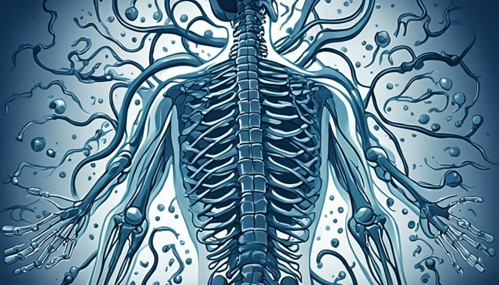
Knowing what syringomyelia is helps spot its signs, find out why it happens, and choose the best treatment. This is vital for those who have this disorder.
Causes of Syringomyelia
Syringomyelia can be caused by a few different things. The main one is Chiari malformation. This is when the brain tissue pushes through the hole at the bottom of the skull.
This can happen if you have a spinal cord injury. Or, if a tumor grows on your spinal cord. Infections can also lead to syringomyelia. Sometimes, doctors don't know the exact reason why it happens. But, problems at birth, like tethered cord syndrome, can be at the root of it too.
Chiari Malformation
Chiari malformation often causes syringomyelia. It makes part of your brain go into your spinal canal. This blocks the normal path of cerebrospinal fluid (CSF). A syrinx, or fluid-filled area, then forms in your spinal cord.
Spinal Cord Injuries
If you have a traumatic injury to your spinal cord, it might lead to syringomyelia. This injury can disturb the CSF's flow, creating a syrinx. What's surprising is, this might happen years after the original injury.
Tumors and Growths
Spinal cord tumors, even non-cancerous ones, can block CSF from moving freely. This blockage can cause a syrinx. There are other types of growths too, like arachnoid cysts, that can prompt syringomyelia.
Congenital Abnormalities
Certain birth defects can also be a reason for syringomyelia. For example, tethered cord syndrome. This is when your spinal cord is fixed too tightly. It can stop CSF from flowing right, creating a syrinx.
Diagnosing Syringomyelia
Diagnosing syringomyelia involves reviewing your medical history and a neurological exam. It also includes advanced imaging tests. These steps are vital to find out if a fluid-filled cyst is in your spinal cord.
Medical History and Physical Examination
Your doctor will ask about your past health, looking for signs of syringomyelia. They will test your reflexes, strength, and how you feel and move. This helps them see if there are any nerve problems.
Imaging Tests
For a definite diagnosis, an MRI is the best tool. This scan shows your spine and can find a syrinx. It can also show if there are serious issues like Chiari malformation or tumors.
Occasionally, a dynamic MRI is needed to see how fluid around the spine moves. Sometimes a dye is used to get clearer pictures.
Combining your history, exam, and imaging results helps the doctor know if you have syringomyelia. This leads to a treatment plan designed for your situation.
Treatment Options for Syringomyelia
The way syringomyelia is treated depends on how bad the symptoms are and if they're getting worse. If the condition doesn't show symptoms, your doctor might just check on you regularly without any specific treatment at first.
Monitoring and Observation
If your syringomyelia isn't causing a lot of issues, the plan might include getting regular MRIs. This way, doctors can keep an eye on the syrinx's size and location. This monitoring helps ensure your health stays steady.
Your healthcare team will decide how often you need to come in and have imaging tests. They will make sure the illness isn't getting worse.
Surgical Interventions
When syringomyelia brings bad or worsening symptoms, surgery might be the best step. The main aim of surgery is to get rid of the syrinx to stop more spinal cord damage. Surgery might work by:
- Fixing a Chiari malformation to help CSF flow normally
- Stop a syrinx from getting bigger after a spinal cord injury
- Take out things like scar tissue or tumors that block CSF flow
- Drain the syrinx directly using a shunt or other methods
Which surgery you have depends on what caused your syringomyelia, the syrinx's size and place, and your general health. Your neurosurgeon will choose the best surgery for you closely.
Even after successful surgery, syringomyelia could come back. So, seeing your doctor regularly and possibly more treatment are important. Some symptoms could stay because of damage to the spinal cord and nerves after treatment.
Living with Syringomyelia
Managing syringomyelia means handling symptoms and adjusting your lifestyle. This is key for a good quality of life. It includes using pain meds, doing physical therapy, and using occupational therapy. They help with muscle weakness, feeling loss, and neurological issues.
Managing Symptoms
Dealing with pain is vital in this condition. A doctor may give you medicine to ease the chronic pain. Physical and occupational therapy are also great. They improve muscle function, coordination, and daily tasks.
Lifestyle Adjustments
Making some changes can help in managing syringomyelia. Try to avoid heavy lifting to reduce symptom risks. Keeping a healthy weight, staying active, and lowering stress can improve your well-being.
Support Resources
Joining support groups and reaching out for help is important. The American Syringomyelia & Chiari Alliance Project (ASAP) can offer great support. They provide information, emotional support, and a community. Seeing a neurosurgeon or neurologist regularly is crucial too. They help track the condition and plan the best treatment.

Syringomyelia in Children
Syringomyelia can show up in kids, often with a Chiari malformation or other related issues. It may only lead to scoliosis or other spinal issues in them. Catching it early and treating it is crucial to stop more damage to the spinal cord and brain.
Kids may need special care from a pediatric neurosurgeon or neurologist. They might also need surgery. This helps to fix the problem and handle the syrinx.
Syringomyelia in children may cause weakness, pain, or trouble with reflexes. Finding out if a child has it involves physical exams and looking at their spinal cord with MRI. Shunting the cyst and treating what's causing the problem, like Chiari malformations, are common ways to treat it.
After diagnosis, children need regular check-ups. They might also need physical or occupational therapy. While it's not common, parents and doctors should know about this condition in kids and how to deal with it.
Complications of Syringomyelia
Syringomyelia is a disorder where a fluid-filled cyst forms within the spinal cord. It can cause many problems if the syrinx grows or hurts the spinal cord's nerves. These include:
- Scoliosis: A sideways curve of the spine, can happen with this disorder.
- Chronic pain: The syrinx can damage the spinal cord, leading to long-term, intense pain.
- Motor difficulties: It can cause leg muscles to weaken and stiffen, making walking hard.
- Paralysis: In very severe cases, it may lead to paralysis if nerve damage occurs.
The complications you face depend on the syrinx's size and location, and what caused it. Getting diagnosed early and treated well is key to avoiding or dealing with these issues. This approach can greatly help manage your quality of life.
Syringomyelia and Associated Conditions
Syringomyelia often comes with other health issues, with Chiari malformation being very common. In Chiari malformation, the brain stretches into the spinal canal. This blocks the usual flow of cerebrospinal fluid (CSF) and causes a syrinx, which is a fluid-filled cyst in the spinal cord.
Syringomyelia can also be linked to spinal cord injuries, meningitis, arachnoiditis, tethered cord syndrome, and spinal cord tumors. It's key to understand how syringomyelia and these conditions are connected. This helps in diagnosing it right and finding the best treatment. It also helps in managing one's health when living with this nerve disorder.
In some cases, spinal cord injuries lead to syringomyelia later on, even years after the injury. Conditions from birth, like tethered cord syndrome, might also cause syringomyelia to show up when people are between 25 and 40 years old.
Finding the connection between syringomyelia and these health problems is vital for doctors. It helps in making the right diagnosis and providing the best care. This understanding can make life better for those dealing with syringomyelia and its effects.
Research and Advancements
Research is ongoing to understand and treat syringomyelia better. The National Institute of Neurological Disorders and Stroke (NINDS) supports a lot of this work. It focuses on figuring out how genetics play a part in Chiari malformation, which often leads to syringomyelia. It also looks to make better ways to see inside the body, improve treatments, and prevent cell damage in the spine that causes this condition.
Clinical trials play a big role in this research. They help scientists learn more about syringomyelia and try new treatments. Participating in these trials gives patients a chance to help move research forward and improve care for everyone dealing with syringomyelia. Thanks to efforts on genetics, better tests, and new treatments, the outlook for those with syringomyelia is improving.
Prevention of Syringomyelia
Syringomyelia is a condition that has many causes, like Chiari malformation and spinal cord injuries. While not all causes can be prevented, there are steps to lower the risk. It’s crucial to manage Chiari malformation properly and treat any other conditions early. This care can help prevent syringomyelia. For spinal cord injuries, getting help soon and going through rehab can stop syrinx from forming.
Taking folic acid as a supplement during pregnancy is another way to lower risks. This helps prevent birth defects linked to syringomyelia prevention. Staying in touch with your doctors regularly if you have risks for syringomyelia is also very important.
Conclusion
Syringomyelia is a complex neurological disorder. It forms a syrinx, a fluid-filled cyst, in the spinal cord. This can cause pain, weakness, and loss of feeling.
Different factors can lead to syringomyelia. The most common is the Chiari malformation. It blocks the flow of cerebrospinal fluid.
It's key to diagnose syringomyelia accurately. This is done through medical history, exams, and imaging tests. Treatment can involve watching closely, surgery, or both.
The National Institute of Neurological Disorders and Stroke (NINDS) funds a lot of the research on syringomyelia. Ongoing studies aim to understand and treat it better. This helps both patients and doctors make informed choices.
FAQ
What is syringomyelia?
Syringomyelia is a rare neurological disorder. It causes a fluid-filled cyst to form in the spinal cord. This cyst can damage the spinal cord and press on nerve fibers. These fibers send information to and from the brain.
What are the types of syringomyelia?
There are two main types of syringomyelia. Congenital syringomyelia is one type. Acquired syringomyelia is the other. These types also go by the names communicating and noncommunicating syringomyelia.
How common is syringomyelia?
Syringomyelia is quite rare. It affects about 8 out of 100,000 people in the U.S. Some risk factors include Chiari malformation or spinal cord injuries.
What are the symptoms of syringomyelia?
At first, you might feel pain and weakness. You could also have stiffness and headaches. Later, symptoms might include problems with bodily functions and spine curvature.
What causes syringomyelia?
A Chiari malformation is the main cause. It happens when brain tissue extends into the spinal canal. This blocks the flow of fluid. There are other causes too, like spinal cord injuries and tumors.
How is syringomyelia diagnosed?
Doctors use your medical history, a physical exam, and imaging tests. An MRI is especially important. It shows if you have a syrinx and where it’s located.
How is syringomyelia treated?
Treatment depends on your symptoms and how fast they’re getting worse. If the condition isn’t serious, doctors might just watch it. But, if it’s bad or getting worse, surgery could be needed to remove the syrinx.
How can people with syringomyelia manage their condition?
Managing your condition means dealing with symptoms and making lifestyle changes. This could include taking pain meds, doing physical therapy, and avoiding things that make you feel worse. Support groups and resources can also help a lot.
Can children develop syringomyelia?
Yes, children can get syringomyelia. It’s often linked with a Chiari malformation or other birth defects. It’s key to find and treat it early to avoid more damage.
What are the potential complications of syringomyelia?
This condition can lead to scoliosis, ongoing pain, walking issues, and in severe cases, not being able to move. The complications depend on where the syrinx is and what causes it.
Source Links
- https://www.mayoclinic.org/diseases-conditions/syringomyelia/symptoms-causes/syc-20354771
- https://www.ninds.nih.gov/health-information/disorders/syringomyelia
- https://rarediseases.org/rare-diseases/syringomyelia/
- https://www.mayoclinic.org/diseases-conditions/syringomyelia/diagnosis-treatment/drc-20354775
- https://www.nyp.org/ochspine/syringomyelia/treatment
- https://www.seattlechildrens.org/conditions/syringomyelia/
- https://now.aapmr.org/pediatric-syringomyelia/
- https://www.ncbi.nlm.nih.gov/pmc/articles/PMC7276219/
- https://www.ncbi.nlm.nih.gov/books/NBK537110/
- https://www.sciencedirect.com/topics/medicine-and-dentistry/syringomyelia


