Neuroradiology Meaning
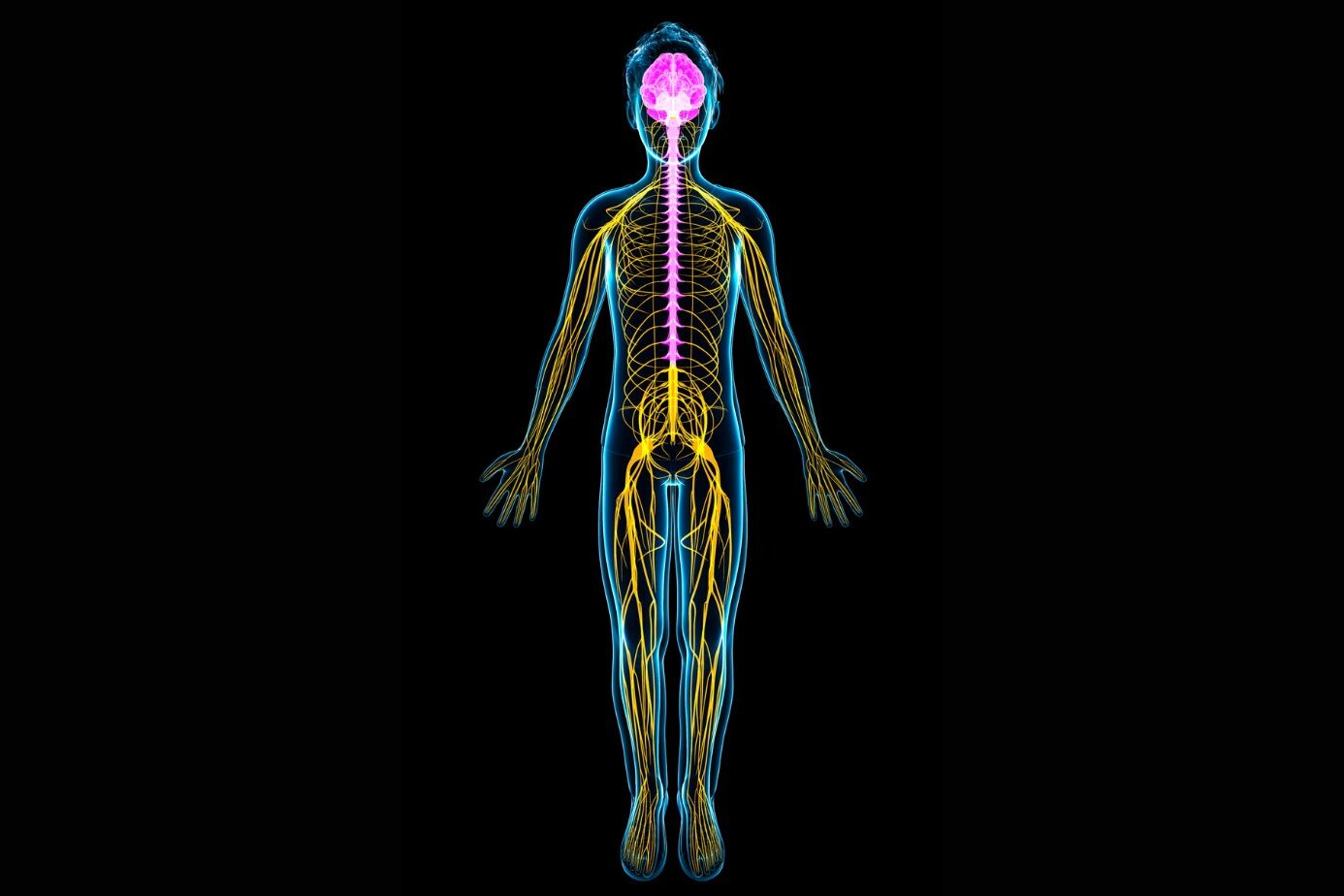
The stellar progress made by medical science over the years has enabled doctors with new technologies. These help them to diagnose and treat conditions, which were once considered implausible. Neuroradiology is one such technology that plays a key role in treating conditions of the nervous system. The nervous system is the most important part of the human body, and any harm caused to it can be life-threatening for the person. Thus, the introduction of neuroradiology has helped in the quick diagnosis and treatment of neurological conditions. It is a subspecialty of radiology that uses neuroimaging techniques to diagnose and treat the abnormalities found in the brain, spinal cord and neck. Tumours, strokes, aneurysms, arteriovenous malformations, etc are treated with the help of neuroradiology. Consult a neurologist in Agra or a brain doctor in Patna for the treatment of these disorders.
Who are neuroradiologists?
Neuroradiologists are neuroradiology doctors- highly skilled professionals, trained to interpret X-rays, CT scans, MRIs. They work with other physicians such as neurologists, neurosurgeons, etc and give insights on the condition of the patient and recommend any further scans or treatments. They are certified professionals who, unlike other radiologists can treat the abnormalities of the central and peripheral nervous system and treat the diseases using minimally invasive, image guided surgery. The best neurologist in Gwalior and neuroradiologists help in treating the abnormalities of the brain.
How does Neuroradiology work?
Neuroradiology uses imaging techniques such as X-rays, Computed Tomography (CT scans of the brain), Magnetic Resonance Image, and ultrasounds. Machines such as magnetic resonance scanner, CT scanner, and ultrasound machines, help in producing these images. A neuroradiologist examines and identifies the characteristics of the images. Based on their knowledge and experience, neuroradiologists identify the problems the patient has. With the help of the inputs given by neuroradiologists, the physician in concern treats the patient. You may consult the best neurologist in Patna or get online neurologist consultation. Some of the techniques involved in neuroradiology are –
- X-Rays – X-rays or X-radiations are the most common type of medical imaging techniques. They use a small amount of radiation to penetrate through your body and create black and white images of the body’s internal structure. The radiation dose depends upon the type of exam and the area of the body to be examined.
- CT methods – CT scans are computerized imaging techniques, where a narrow beam of x-rays is aimed at the patient and quickly rotated all around the body. A computer then helps in assembling the images in a multidimensional view. CT scans help in procuring detailed images of the body by using non invasive methods. Images of the blood vessels, soft tissues, organs, etc can be viewed more clearly with the help of CT images. These are very helpful in diagnosing spinal pain, brain tumours or internal bleeding.
- Diagnostic MRI methods – MRIs are yet another diagnostic imaging technique that uses radio waves, and strong magnetic fields to get cross-sectional images of your internal organs.
- Ultrasound – An ultrasound or sonography is an imaging technique that uses sound waves to get images of the internal parts of the body. A gel is placed on part of the body to be examined and an external probe is used to transmit and collect the sound waves. Ultrasounds are helpful to diagnose the causes of pain, swelling, or infection on the inside of the body.
- Catheter angiography – Here, a thin plastic tube, a catheter is inserted into an artery. A radiopaque material is injected through the tube to capture images.
- Embolization – Embolization is a minimally invasive technique, used by interventional neurologists, which helps in blocking the blood supply to blood vessels which may cause harm to the person.
- Coil placement – Coiling, also known as endovascular coiling is a minimally invasive technique that treats an aneurysm. Coiling is done by placing a thin wire-a coil to stop the blood flow, thus preventing ruptures. A catheter is inserted in a groin artery and then is advanced to the affected brain artery. The coil is dyed so that it can be guided by the X-rays to reach the affected part and deploy the coil there. A coil is usually made up of metal.
- Stent placement – Here, catheters are used to unblock the blood vessels and improve the blood flow in vessels of the neck and the brain. The doctor will place a balloon-tipped catheter, as guided by x-ray into an artery or vein. The balloon is then inflated and deflated to stretch the artery wall, thus restoring blockage.
Benefits of Neuroradiology
The discovery of neuroradiology has been a gift to the medical sciences. It has facilitated surgeons in the quick and early diagnosis of illnesses. Due to the expeditious imaging techniques, the need for exploratory surgery has been eliminated. Neuroradiologists can get prompt images of the deep interior parts of the brain and the spinal cord, and determine if there’s a need for surgery. With the help of interventional radiology, doctors can use image-guided, 3D technology to treat conditions, which are not only efficient but also minimally invasive. The risk of damage is low and the recovery is speedy, by the virtue of neuroradiology. Consult the famous neurologist in Patna and the best neurologist in Gwalior through online neurologist consultation or neurologist online chat for the treatment of your neurological disorders.
About The Author

Medically reviewed by Dr. Chandril Chugh, MD, DM (Neurology)
Dr. Chandril Chugh is a U.S.-trained, board-certified neurologist with expertise in diagnosing and managing neurological disorders, including migraines, epilepsy, Parkinson’s disease, and movement disorders. His clinical focus includes evidence-based neurological care and patient education.
All content is reviewed for medical accuracy and aligned with current neurological guidelines.