
Breathing Exercises For Anxiety: Easy Techniques To Calm Your Mind
Breathing exercises for anxiety give you a fast, practical way to tell your nervous system that the…

Natural Ways To Reduce Anxiety: Key Methods & Simple Strategies
Natural ways to reduce anxiety give you practical tools to calm your mind and body without relying…
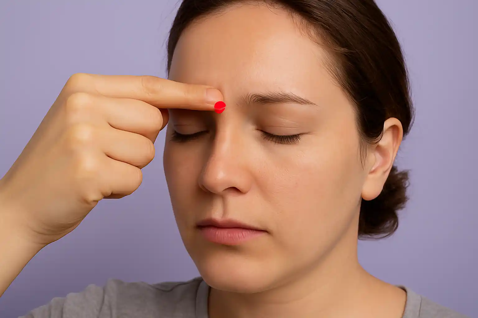
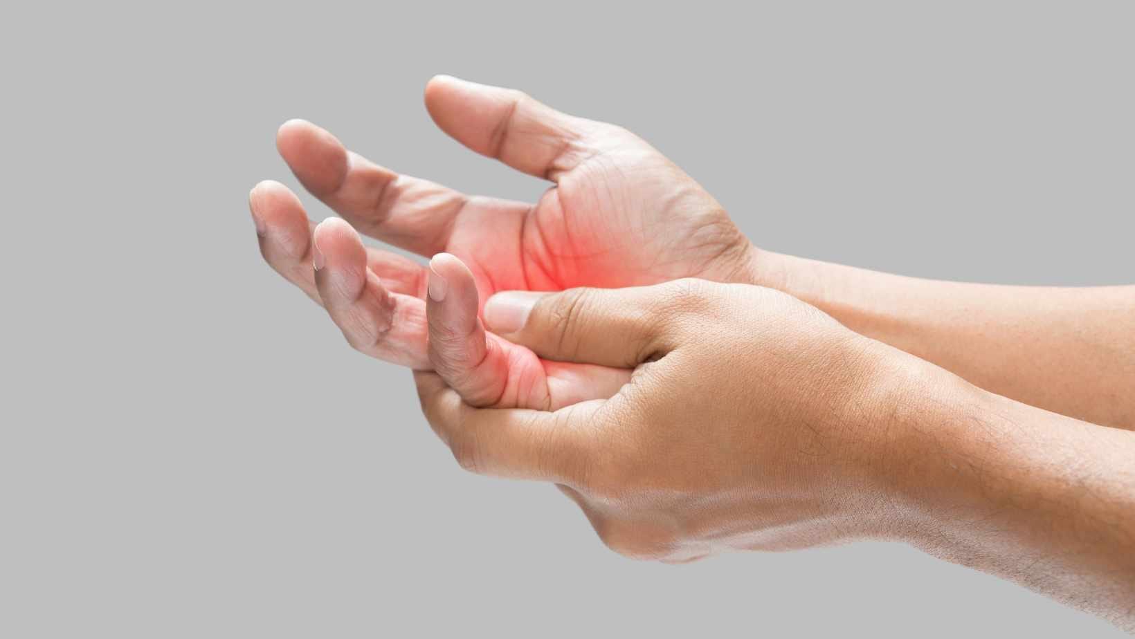
Pressure Points For Anxiety Relief: How Acupressure Works
Pressure points for anxiety are small spots on your skin that respond strongly to firm touch. When…

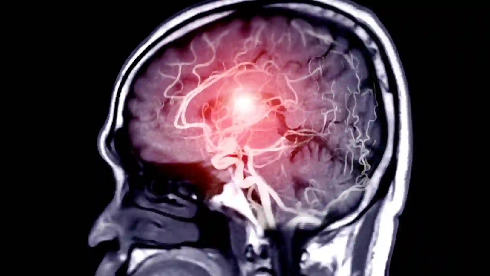
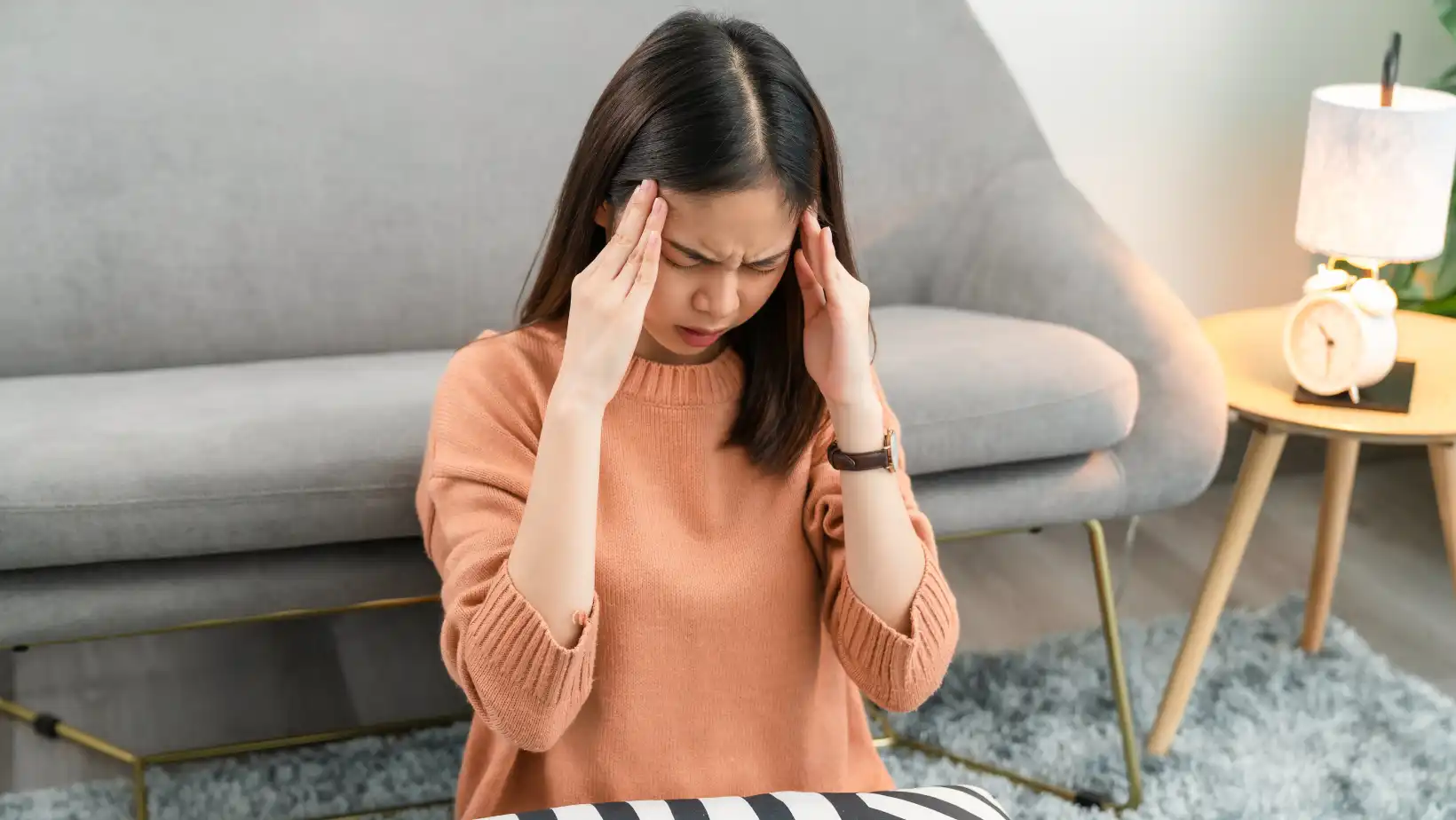
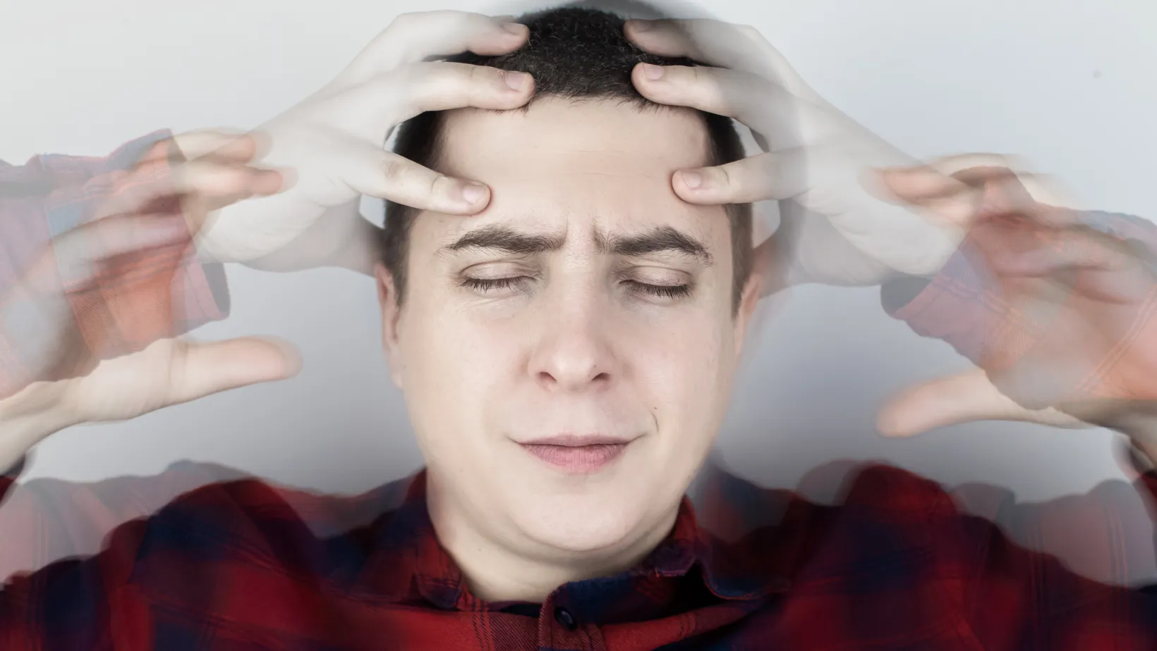
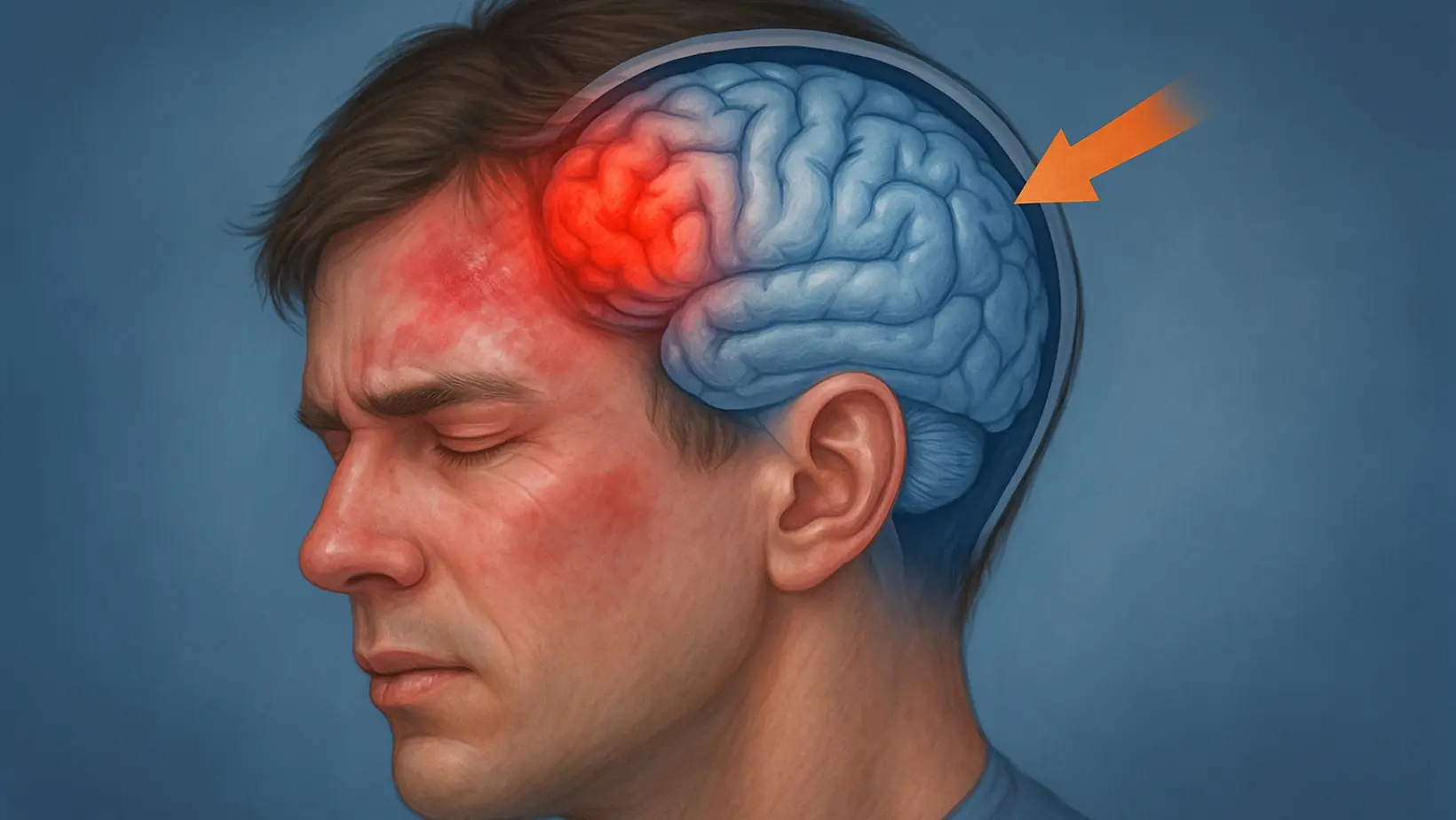
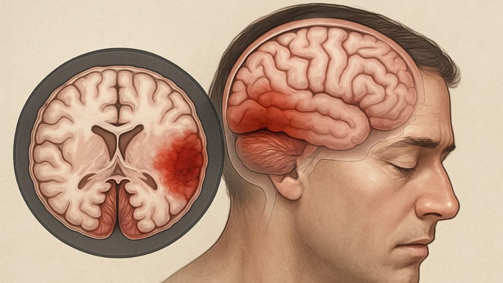
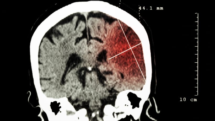
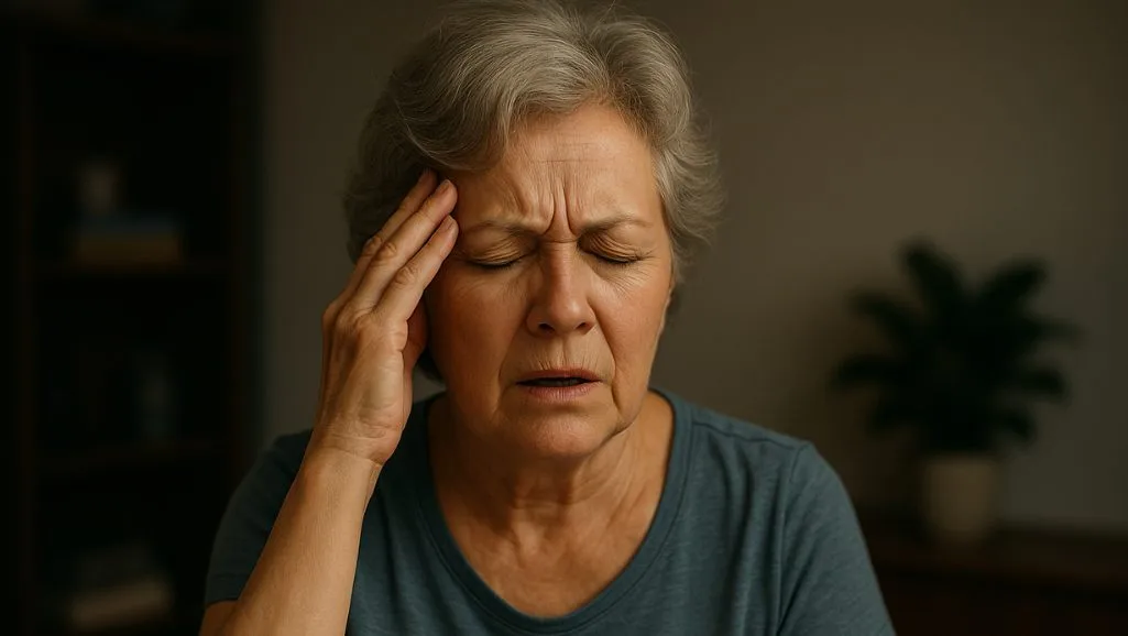
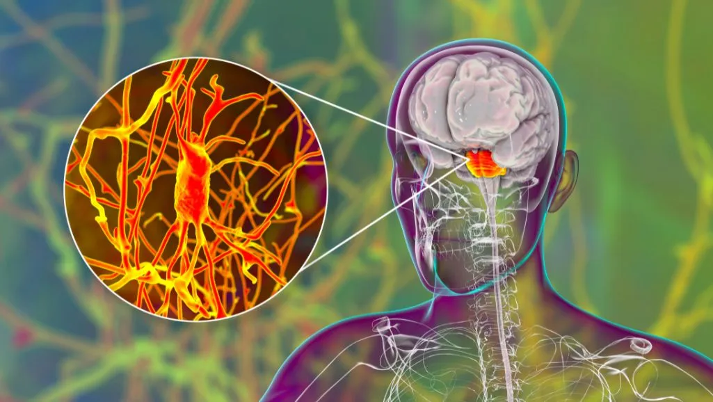
Migraine vs Stroke: Signs, Symptoms & When to Act
Migraines and strokes both affect your brain, and the warning signs can look alike. Doctors warn…

How Long Does Vertigo Last? Duration, Recovery & When to Seek Care
Vertigo can last seconds, minutes, hours, or even days, depending on the cause. Most short episodes…

Bell’s Palsy vs Stroke: Key Differences, Symptoms & Diagnosis
Bell's palsy and stroke can cause a drooping face on one side. Yet the damage inside your body is…

Can You Die From A Stroke? Causes, Risks, Fatal Signs & Survival Outlook
Stroke is one of the top causes of death in the world and in the United States. Global data from…
Stroke vs Aneurysm: Key Differences & Symptoms
A stroke happens when blood flow to part of the brain stops or when a vessel bursts and bleeds. An…

What Is Stroke-Level Blood Pressure? Numbers, Risks & Emergency Signs
Stroke-level blood pressure means your blood pressure is so high that your brain, heart, and other…

Can Stress Cause A Stroke? Causes, Symptoms & Prevention
Long-term stress can raise blood pressure, disturb sleep, change hormones, and increase…

Can Stress Cause Vertigo? Causes, Symptoms & Care
Vertigo means a false sense that you or the room is spinning when nothing is moving. Dizziness can…

Can Stress Kill You? How Stress Damages The Body
Stress does not usually stop the heart by itself. But strong and long-lasting stress can raise the…

Can Stress Cause Nosebleeds? Mechanisms & Risks
Stress does not usually act as the only cause. Yet it can push your body in ways that make bleeding…

How I Cured My Sjögren’s Syndrome: Natural & Medical Insights
Sjögren’s syndrome is an autoimmune disease. Your immune cells attack moisture glands and sometimes…

Importance Of Mental Health Awareness: Why It Matters
The importance of mental health awareness starts with a simple fact. Your mind health shapes how…
Natural Remedies for Nerve Pain: Home Treatments & Herbal Relief
Natural remedies for nerve pain help you calm burning, tingling, and pins and needles without heavy…
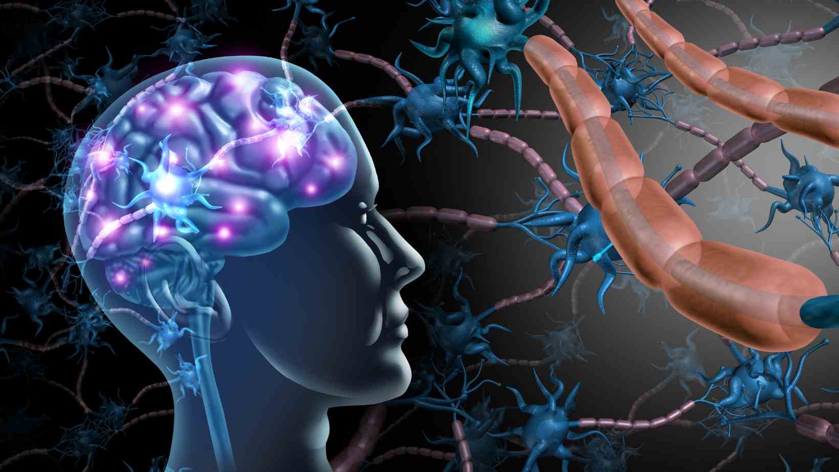
Effects of Energy Drinks on the Brain: What Science Reveals
The effects of energy drinks on the brain are immediate and lasting. You feel more awake and…

Mental Health Activities for Adults: Games, Exercises & Self-Care Ideas
Mental health activities for adults are daily actions that strengthen emotional balance, lower…

Child Development Psychology: Stages, Theories & Growth
Child development psychology explains how you grow from before birth to your teenage years. It…

Feeling Jittery: Common Causes and How to Calm Down
Feeling jittery means your body feels shaky, wired, or on edge. You may notice trembling hands, a…

Best Energy Drinks for Focus, Workout, and Daily Use
Best energy drinks give you focus, steady energy, and simple labels. You want clean caffeine, smart…

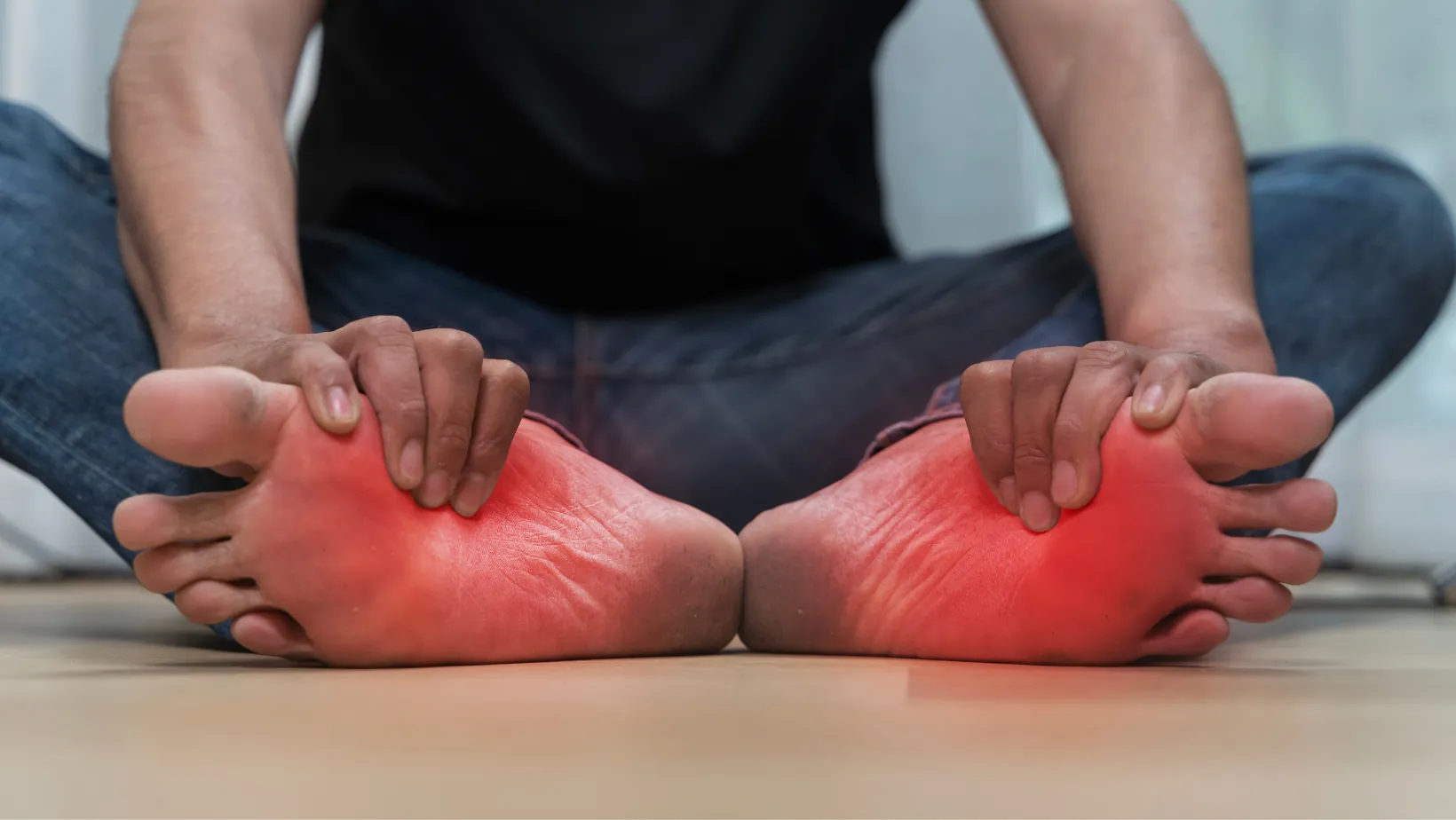
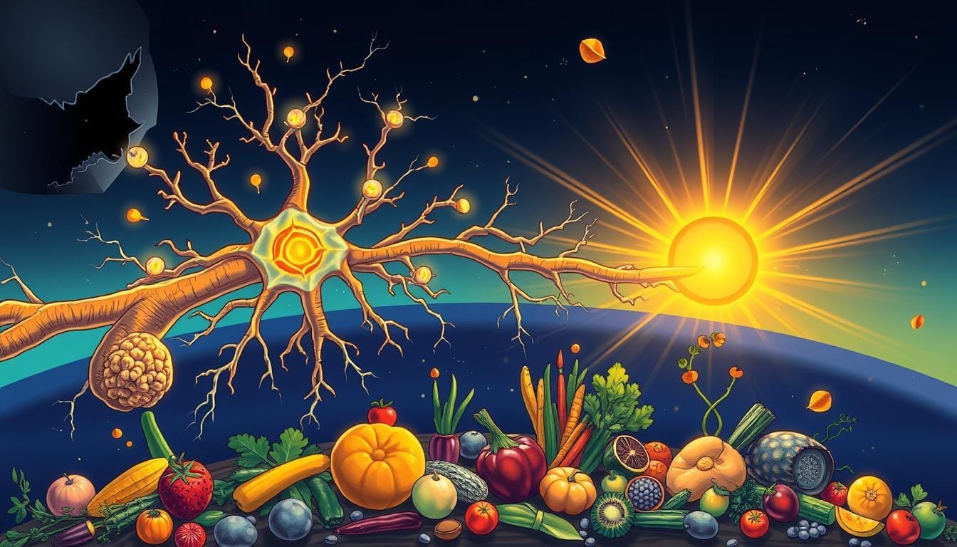
Best Foods for Nerve Repair and Regeneration
You can improve nerve repair by eating the right foods. Good meals give vitamins, healthy fats, and…

ADHD Waiting Mode: What It Is and How to Break Free
ADHD waiting mode happens when you know you have something coming up (a meeting, deadline, or…
Foods That Trigger Migraines (and What to Eat Instead)
Foods that trigger migraines are more common than most people think. For many migraine sufferers,…

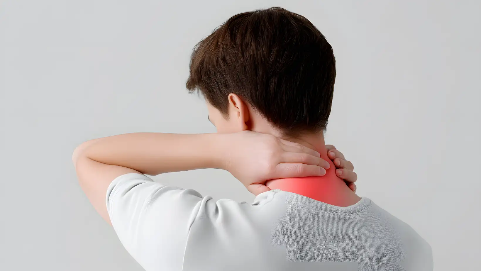
Sinus Neck Pain: Causes, Symptoms & How to Relieve It Naturally
sinus neck pain often surprises people. It starts in your sinuses (the hollow spaces around your…

Waking Up With Neck Pain: Why It Happens and How to Fix It
Waking up with neck pain is one of the most common complaints among adults today. It can start as a…
Neck Pain: Causes, Symptoms, and Warning Signs You Shouldn’t Ignore
Neck pain causes and symptoms can affect anyone at any age. When your neck aches, it is not simply…
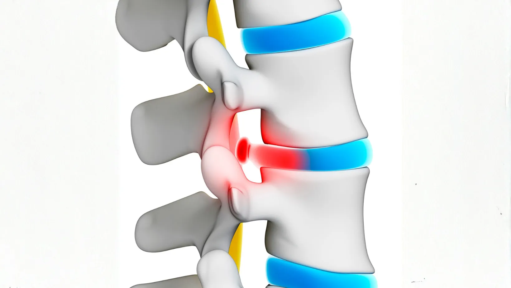
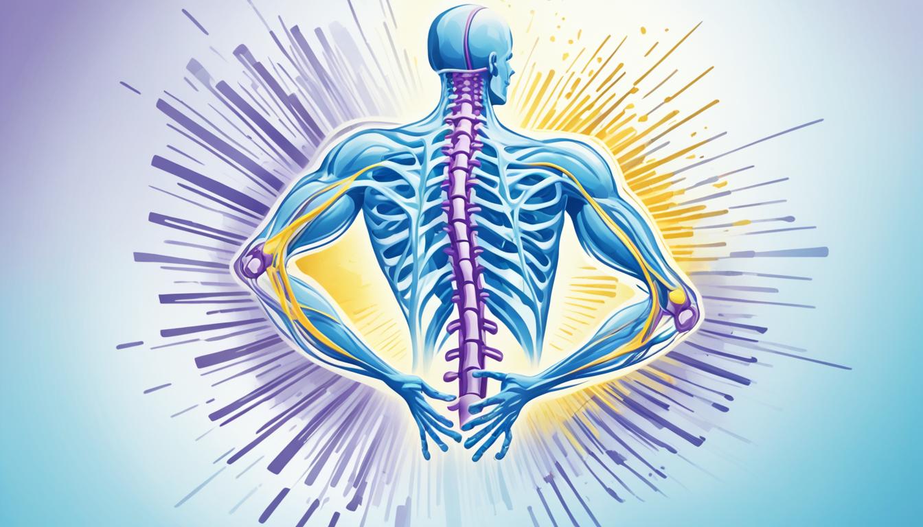
How to Heal a Bulging Disc Naturally: Treatments, Exercises & Recovery Tips
A bulging disc occurs when one of the soft cushions (called intervertebral discs) between your…

What’s The Link Between PTSD and Migraines in Veterans?
PTSD and migraines often appear together in veterans, raising questions about why trauma in war…

Massage Therapy for Migraines
Massage therapy for migraines is gaining attention as a safe and effective way to reduce the…

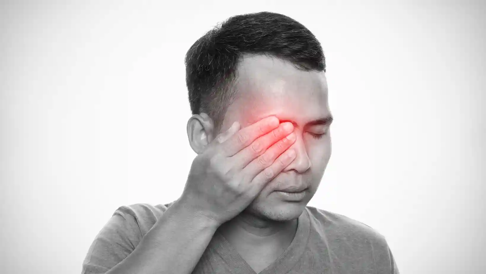
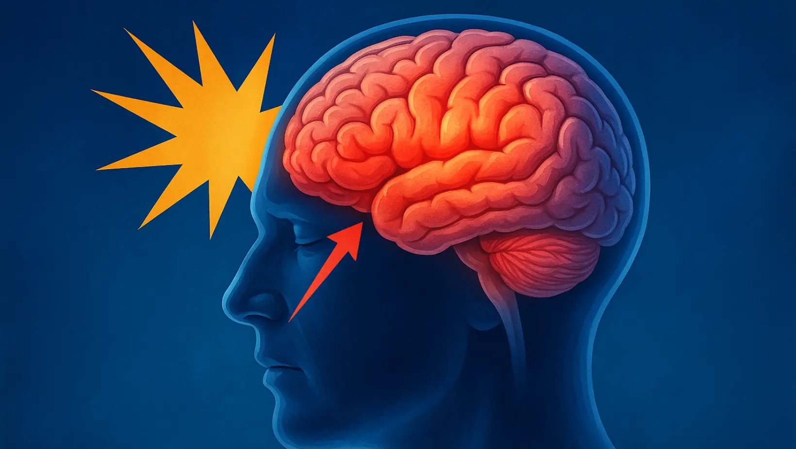
Why Do You Have Migraine Eye Pain?
Migraine eye pain is one of the most troubling symptoms for many people living with migraines. It…

Why is Migraine More Common in Women vs. Men?
Migraine in women vs men has been a mystery for decades. Research indicates that women experience…
Self-Care Tips for Migraine
Migraine self-care is more than just a quick fix. It is about building daily routines, using safe…

What Is Status migrainosus?
Overview Status migrainosus is a rare but serious type of migraine. Unlike a usual migraine that…
What Is Ocular Migraine (Retinal migraine)?
Ocular migraine is a condition that causes temporary vision changes, often frightening the person…

Understanding Types of Migraine
Types of migraines are not all the same. Some strike with flashing lights. Others cause dizziness,…

Migraine vs. Chronic Migraine: Understanding the Difference
episodic vs chronic migraine is a key topic for anyone struggling with frequent headaches. Many…

Chronic Migraine: Causes, Symptoms and Treatment
Chronic migraine is more than just a bad headache. It is a disabling brain condition that affects…
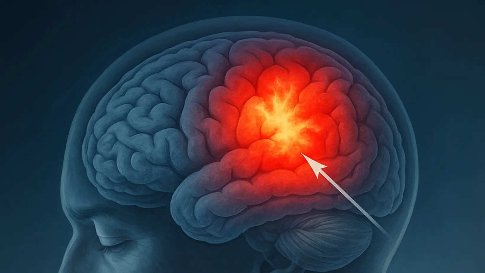
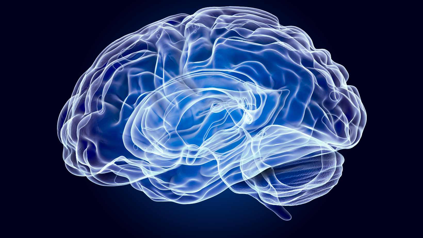
Is There Such a Thing as a ‘Migraine Brain’?
Migraine brain is a term doctors and researchers use to describe the brain of someone who…

Foods and Drinks That May Help Manage Migraine
Foods to prevent migraines play a big role in how often attacks happen. The right meals give your…

What causes migraine in females?
Migraines in females are more common in women than men, and the reasons go far beyond “just…
Concussion Symptoms and Treatment: What You Need to Know
A concussion is one of the most common brain injuries worldwide, yet it’s often misunderstood or…
What Is Encephalitis? A Complete Guide to Brain Inflammation
Encephalitis refers to a serious medical condition characterized by swelling of the brain. When…
Head Injury: Causes, Symptoms, and Treatments Explained
A head injury is more than merely a bump or bruise. It can affect the brain, skull, and scalp,…
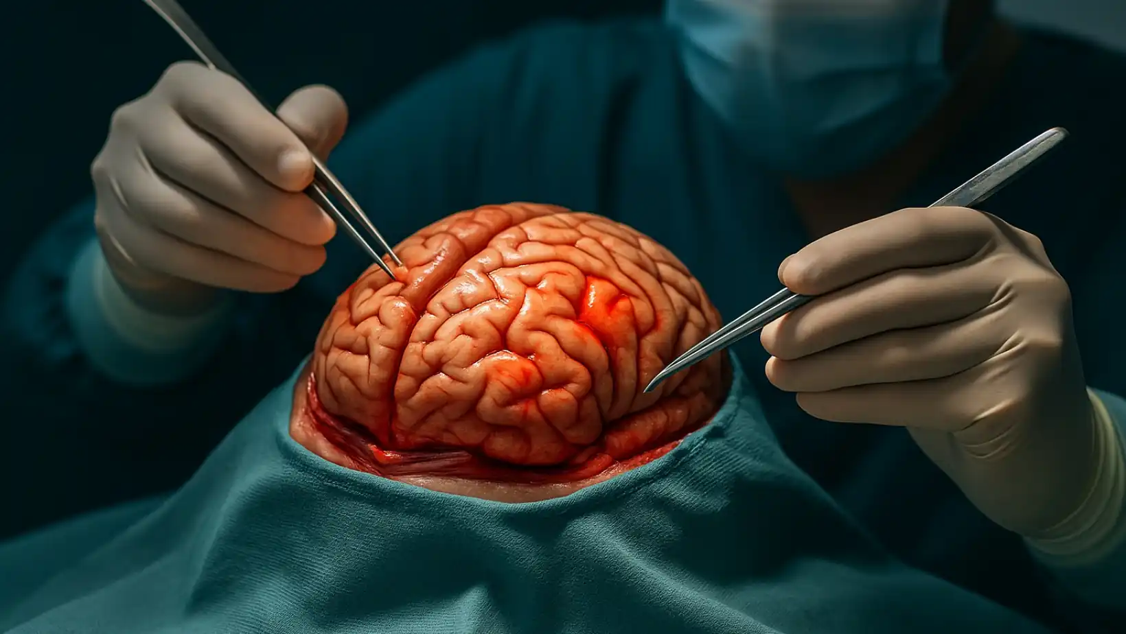
What is Brain Surgery? Understand its Procedures, Benefits & Recovery Time
What is brain surgery? Brain surgery is a medical procedure where a neurosurgeon operates on the…
Understanding Brain Herniation
Brain herniation is one of the deadliest conditions in medicine. It happens when part of the brain…
Brain Disorders: Causes, Types, Risks, and Outlook
What are brain disorders? A brain disorder refers to any condition that negatively impacts the…
Acute Disseminated Encephalomyelitis (ADEM)
Overview What is acute disseminated encephalomyelitis (ADEM)? Acute disseminated encephalomyelitis…
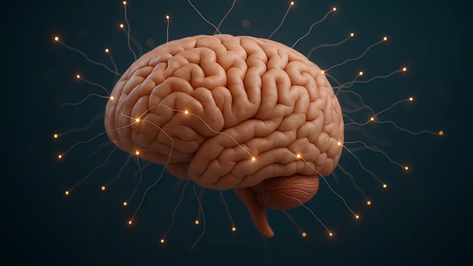
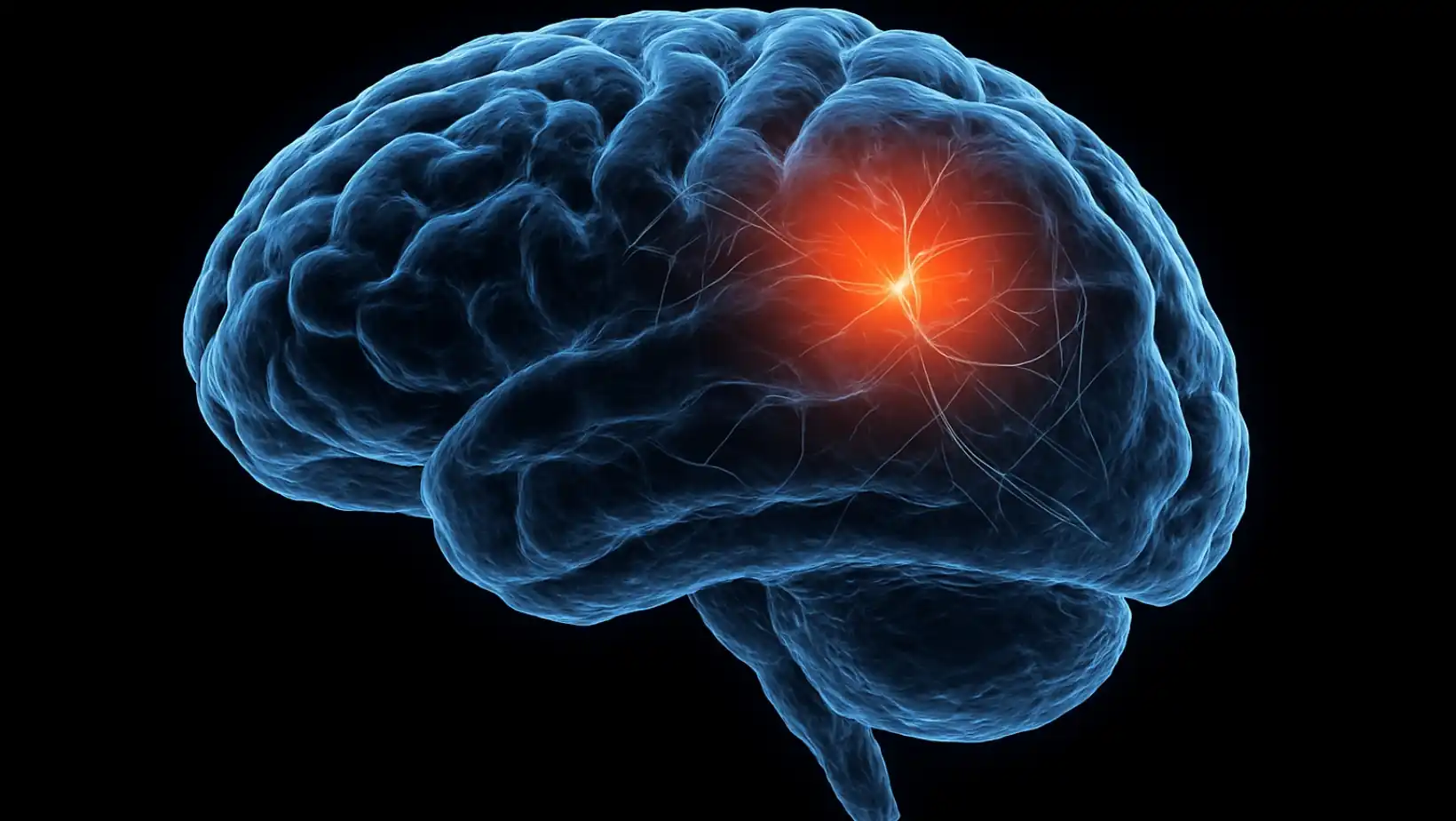
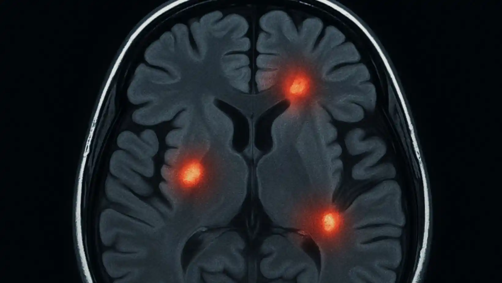
Migraine and Brain Lesions
What Are Brain Lesions? A brain lesion is an area where brain tissue is damaged. The damage can…
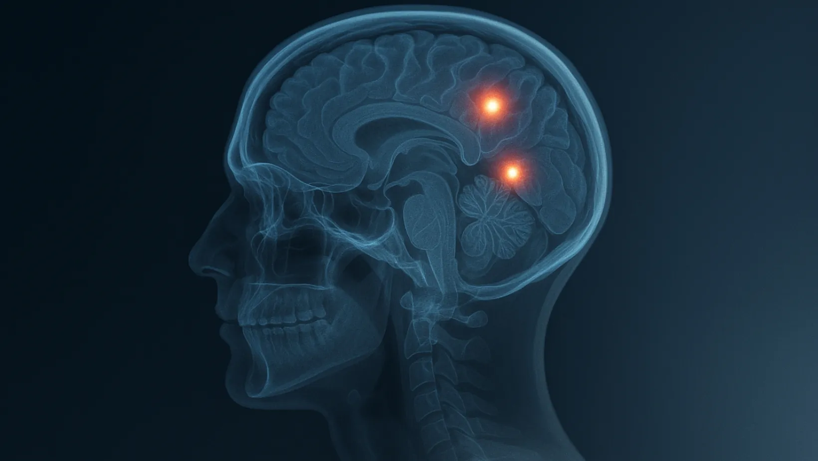
Brain Lesions: Causes, Symptoms, Treatments
Brain lesions are areas of damage in the brain tissue, and understanding them is crucial for your…
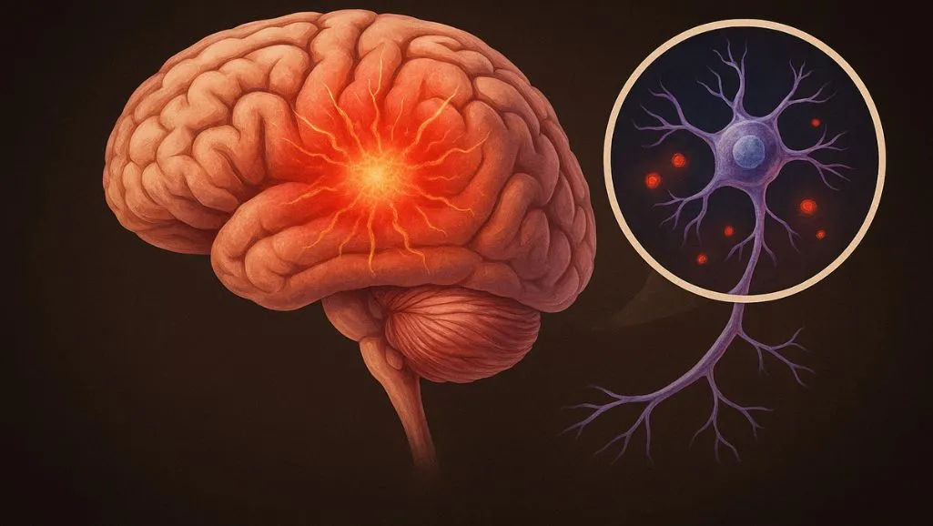
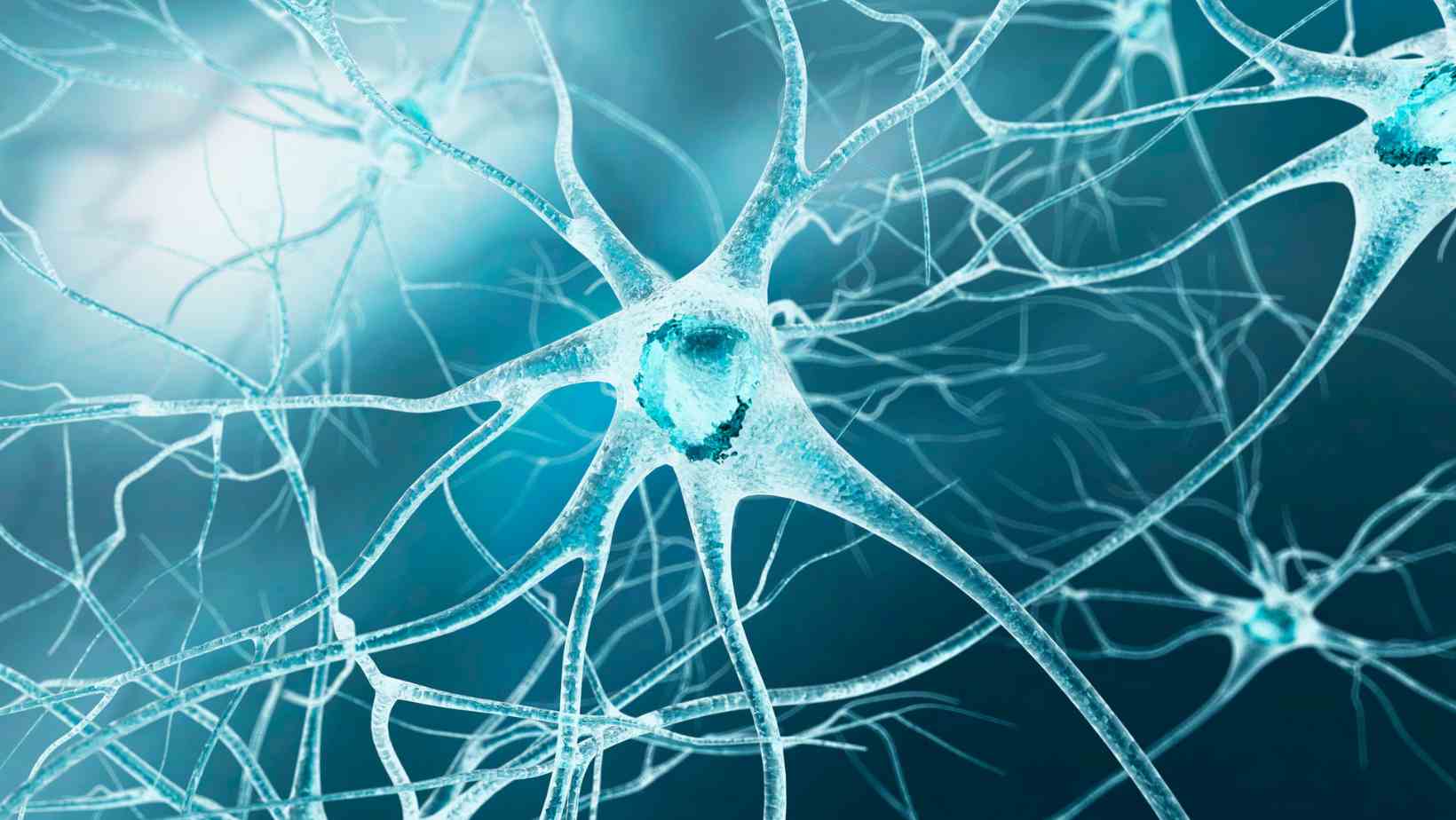
Neuroinflammation Demystified: Triggers, Symptoms, and Treatments
Neuroinflammation is the brain’s defense mechanism. When it detects a threat, like toxins,…
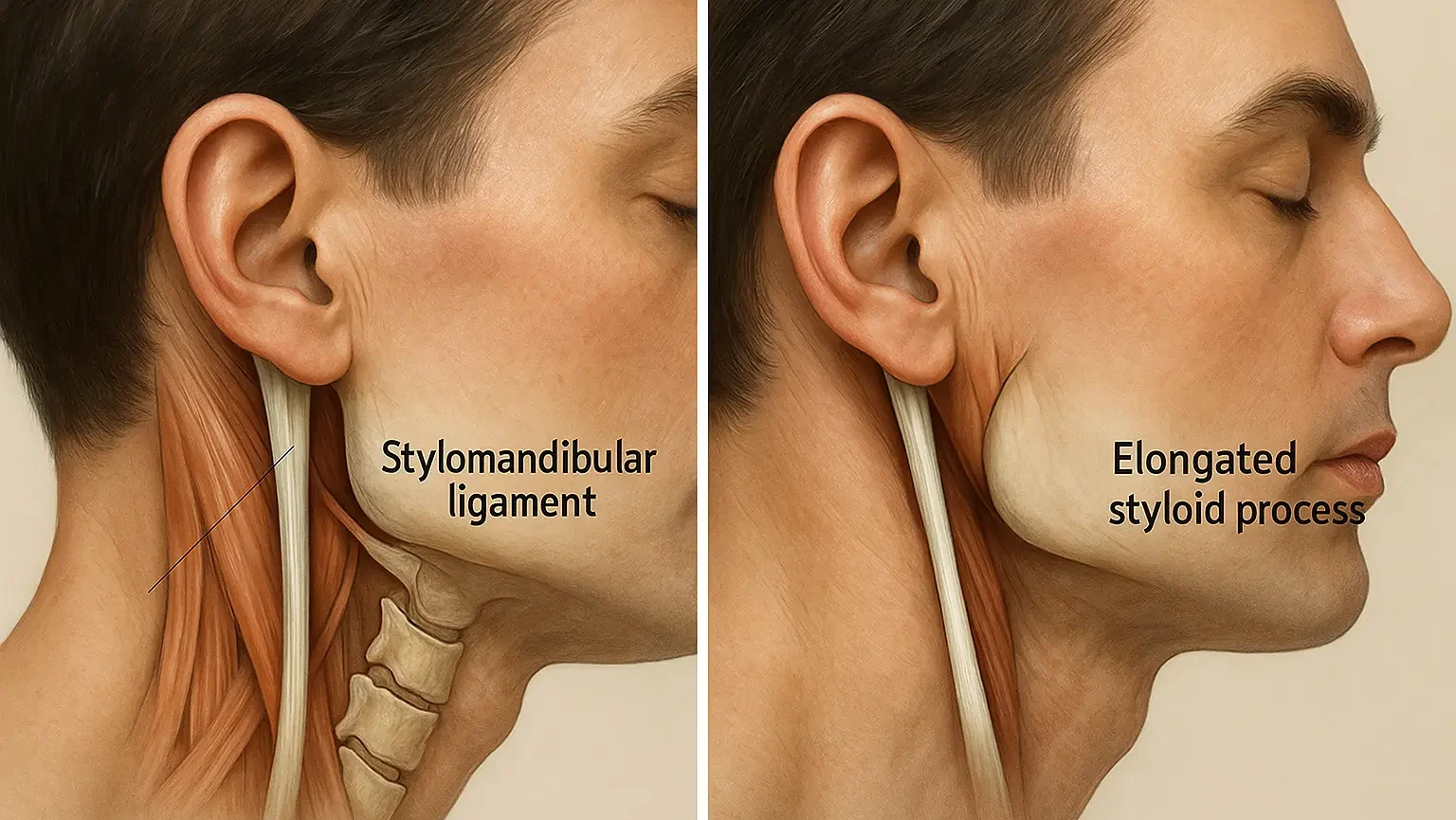
Ernest Syndrome vs. Eagle Syndrome: Differences, Symptoms, Causes, and Treatments
Eagle syndrome and Ernest Syndrome are two rare but painful conditions that affect the head and…
Posterior Reversible Encephalopathy Syndrome (PRES)
Posterior Reversible Encephalopathy Syndrome (PRES) is a brain condition that causes sudden…

What is Uncoordinated Movement?
Uncoordinated movement is when the body struggles to control motion properly. It can affect…
Transient Ischemic Attack (TIA)
Transient Ischemic Attack is a short-term blockage of blood flow to the brain. Though symptoms may…

What is Friedreich’s Ataxia? Understanding the Basics
Friedreich’s ataxia is a rare, inherited condition that quietly alters how the body works. It…
Acute Cerebellar Ataxia (ACA)
Acute cerebellar ataxia doesn’t give you a warning. One day you may walk normally. The next day,…
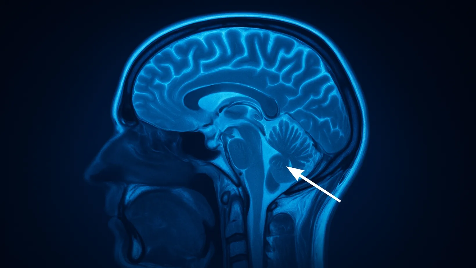
What Is Cerebellar Ataxia? Understanding the Basics
Cerebellar ataxia refers to a condition in which your brain has difficulty managing balance,…

Neurologist-Backed Natural Remedies for Improving Nerve Strength
If you have ever felt tingling, numbness, or pain in your hands or feet, it’s a sign that your…

Superfoods for Brain: Boost Memory & Cognitive Health Naturally
Did you know nearly 60% of your brain is made of fat, and what you eat directly impacts your…
What is Neurosyphilis? Causes, Symptoms & Diagnosis Guide
Have you ever heard about syphilis affecting the brain? That condition is called neurosyphilis, and…

Arnold Chiari Malformation Radiology Insights
Arnold Chiari Malformation (CM) is a condition where brain tissue extends into the spinal canal,…

Masters in Child Psychology: A Complete Career Guide for Future Mental Health Professionals
Did you know that 1 in every 7 children between the ages of 15 and 24 suffers from mental health…

Treatment for Essential Tremor: What Works Best?
Essential tremor is a common neurological condition that causes uncontrollable shaking—usually in…
Effective Brown-Séquard Treatment Options
Ever felt like your body's playing a bizarre trick on you, where one side feels weak while the…

Effective Facial Palsy Treatment Options
Hey there! Ever felt like one side of your face decided to take an unexpected vacation? Maybe a…

The Impact of Smartphone Use on Sleep Patterns
We check our phones before bed, scroll endlessly through social media, and sleep with them under…

Can Stress Cause Brain Tumors? Revealing the Truth Behind the Myth
Ever feel like life's constant pressures – the never-ending to-do lists, looming deadlines, and…

Brain Aneurysms Uncovered: 24 Critical Questions Answered by a Neurologist
As a neurologist, I've stood alongside countless individuals and their families confronting the…

How to Manage Everyday Life with a Neurological Condition
Living with a neurological condition doesn’t mean giving up on your daily life. Yes, the symptoms…

Hereditary Spastic Paraplegia Physical Therapy: How Movement Can Reclaim Life
Hereditary spastic paraplegia (HSP) can make even simple movements feel like a battle. With legs…

Is Sex A Common Cause Of Brain Stroke?
Sex and stroke—two words that rarely appear in the same sentence, yet for some, they intersect in…
Understanding Machado Joseph Disease: Symptoms and Management
Some conditions quietly take root, unfolding their effects over years before a diagnosis is even…
Causes of Mononeuritis Multiplex: Understanding the Underlying Factors
Mononeuritis multiplex is a neurological condition that can affect a person’s quality of life in…
Dandy Walker Syndrome Treatment Options
"Why is my child developing slower than others? Why are their movements not like other kids?"…

Relationship Between Caffeine and Sleep Quality
Caffeine is part of daily life for millions of people across the globe. From the comforting aroma…
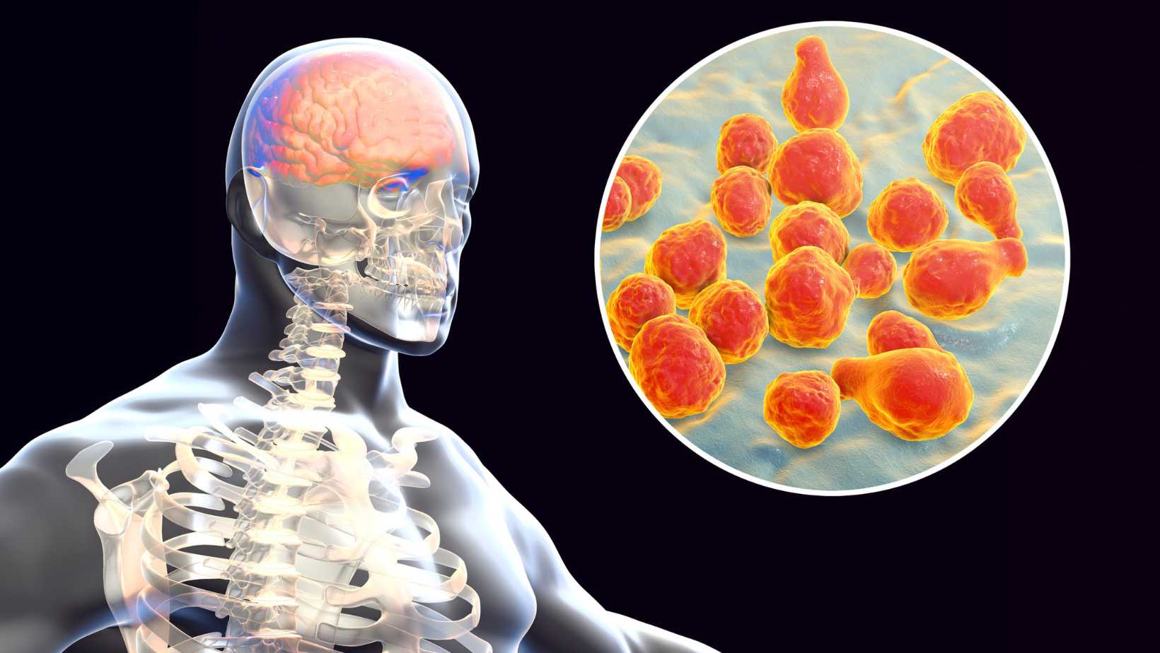
Surgical Management of Meningitis: When is Surgery Needed?
Introduction: Understanding Meningitis and Its Treatment Meningitis is a serious infection that…

The Truth About ADHD Medication in India – Expert Advice by Dr. Chugh
Imagine your mind as a symphony, but the instruments are playing out of tune. That’s ADHD.…
Mind-Body Techniques for Nerve Healing: Stress Reduction & More
Ever feel like your nerves are frayed, buzzing with phantom pain or tingling sensations long after…

Foot Neuropathy and Diabetes: Special Considerations
Imagine your feet as your body's roots, grounding you in the world. Now, picture those roots slowly…

Natural Remedies for Neuropathy: Healing Nerve Pain Without Medication
Say Goodbye to Neuropathy: Natural Treatments That Work Tingling toes? Burning feet? Is that…
Nerve Damage Doesn't Have to Be Forever: Exploring the Possibilities of Repair
Imagine waking up every morning with numb fingers, a burning sensation in your feet, or a sharp,…
Is It Possible to Reverse Neuropathy Fast? What a Neurologist Says
Tired of simply coping with the relentless tingling, numbness, and pain of neuropathy? It’s…

10 Tips for Supporting Nervous System Development in Children
Childhood brain development is a key time for growth and potential. Parents and caregivers can help…
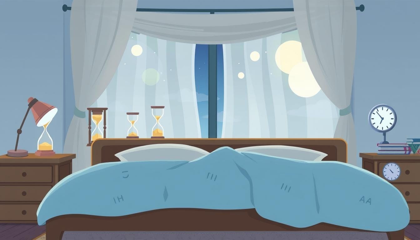
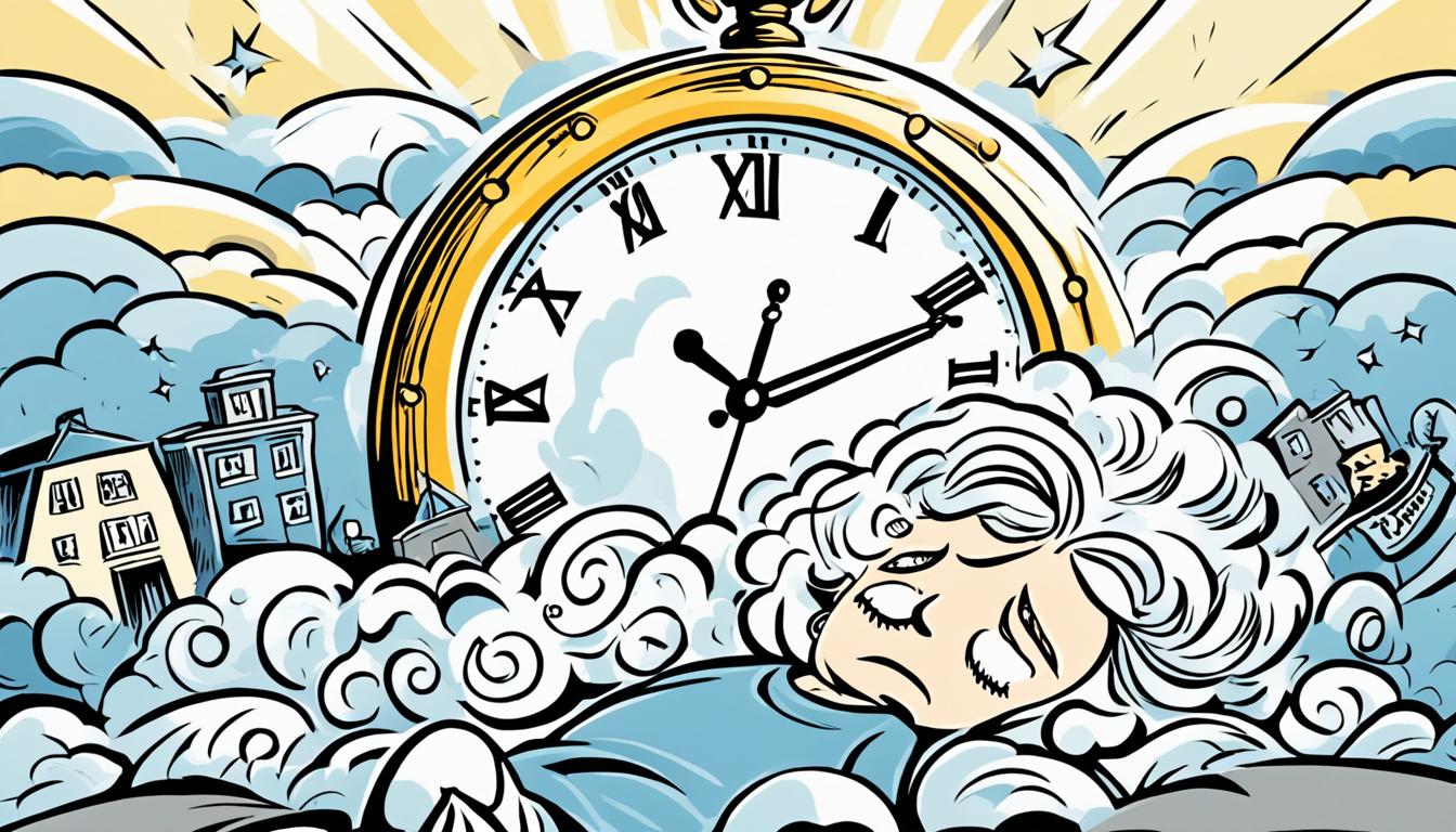
Debunking the 8 Hour Sleep Rule: What You Need to Know
Do you really need 8 hours of sleep every night? Many believe this is a strict rule for good…

How to Create a Bedtime Routine for Adults That Promotes Brain Health
A consistent bedtime routine is key for good brain health and overall well-being. The U.S. Centers…

The Role of Storytelling in Child Development Psychology
Storytelling is a key tool in child development psychology. It helps grow young minds in many ways.…

The Link Between Diet and Childhood Cognitive Growth
Is your child's food helping their brain grow or slowing it down? Parents across the world want…

Foods to Avoid If You Have Neurological Disorders
Neurological disorders can greatly affect a person's health and happiness. Diet is key in managing…
Understanding the Role of Neuroplasticity in Recovery
Have you ever wondered how some people start walking again after a stroke or trauma, even when…
How to Use Nutrition to Support Nerve Regeneration
Peripheral nerve injuries hit over one million people worldwide each year. They cause big problems…

How Energy Drinks Affect Cognitive Performance
Energy drinks have become very popular since the late 1990s. They claim to boost physical…

5 Yoga Poses for Better Sleep and Relaxation
Have you ever spent hours staring at the ceiling, trying to fall asleep but your mind just won't…
Top Methods for Herniated Disc Pain Relief: Effective Solutions
If you're one of the 2% of adults in the U.S. with a herniated disc, you know the pain is real and…
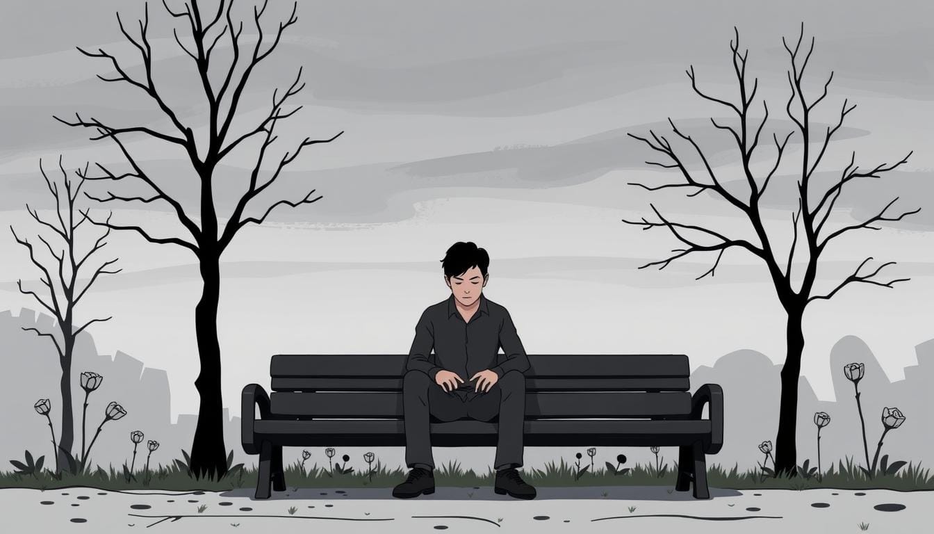
Depression in Men: Symptoms and How to Seek Help | Dr. Chandril Chugh
Depression is a serious mental health issue that affects both men and women. But, depression in men…

Disturbed Mental Health in Older Adults
As the world's population ages, we face a big challenge with the mental health of older adults.…

Top Characteristics of Mental Health: What You Need to Know
Do you ever wonder what makes someone truly mentally healthy? Is it being happy all the time? Is…

Effective Strategies for Bulging Disc Pain Relief
Dealing with a bulging disc can be tough. But, there are ways to find relief and manage the pain.…
Understanding ADHD Insomnia: Causes and Solutions
If you or someone close to you has ADHD, you might know how hard sleep issues can be. ADHD is a…

How to Heal After a Breakup: Tips for Moving On
Breakups can be tough, even if the relationship was ending. Losing your partner means losing shared…